Example 14
Jump to navigation
Jump to search
- Following the 7+2 steps:
- Rhythm
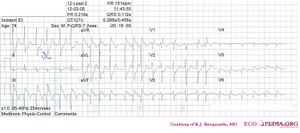
- The ECG shows an irregular rhythm. There are no P waves. Atrial fibrillation.
- Heart rate
- 150 bpm
- Conduction (PQ,QRS,QT)
- PQ: not appropiate QRS: 110ms QT: 360ms QTc: 560ms (prolonged)
- Heartaxis
- QRS positive in I and iso-electric in AVF: horizontal heart axis
- P wave morphology
- No P waves present
- QRS morphology
- Incompleter right bundle branch block. QS V4-V6. Q in III and AVF.
- ST morphology
- ST elevation in III,AVF and V6. ST depression in I, AVL, V2.
- Compare with the old ECG (not available, so skip this step)
- Conclusion?
- Rhythm
Atrial fibrillation with inferior-posterior-lateral myocardial infarction and incomplete right bundle branch block. Lead I shows ST depression, suggestive of right coronary artery involvement.
