Cases and Examples
Jump to navigation
Jump to search
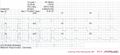
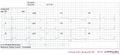
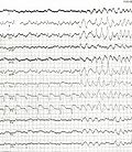
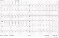
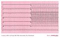
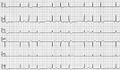
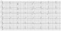
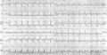
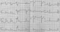
Below you can find some common examples. ECGs can be magnified by clicking on the image....
Ischemia & Myocardial Infarction

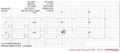
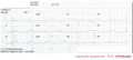
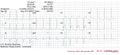
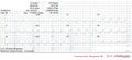
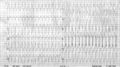
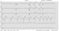
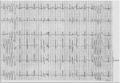
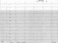
Guess the culprit coronary artery that was occluded in these examples of myocardial infarctions
Examples of Myocardial Infarctions
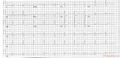
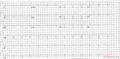
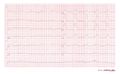
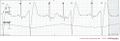
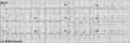
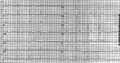
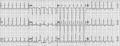
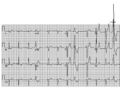
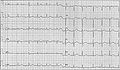
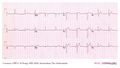
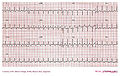
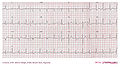
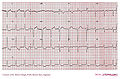
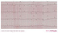
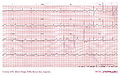
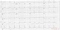 Where is this myocardial infarction located? Case provided by
|
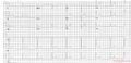
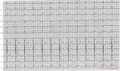
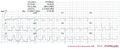
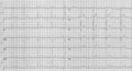
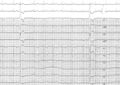
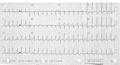
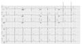
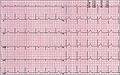
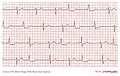
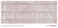
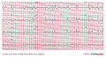
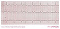
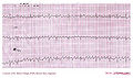
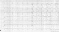 Where is this myocardial infarction located? Case provided by
|
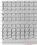
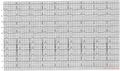
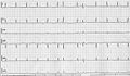
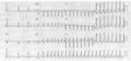
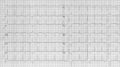
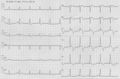
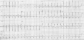
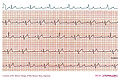
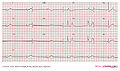
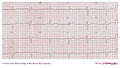
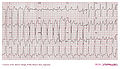
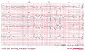
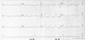 Where is this myocardial infarction located? Case provided by
|
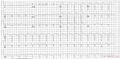
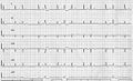
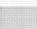
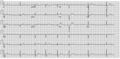
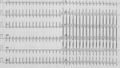
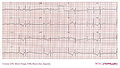
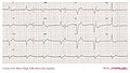
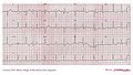
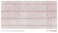
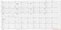 Where is this myocardial infarction located? Case provided by
|
 Where is this myocardial infarction located? Case provided by
|
 Where is this myocardial infarction located? Case provided by
|
 Where is this myocardial infarction located? Case provided by
|
 Where is this myocardial infarction located? Case provided by
|
 Where is this myocardial infarction located? Case provided by
|
 Where is this myocardial infarction located? Case provided by
|
 Where is this myocardial infarction located? Case provided by
|
 Where is this myocardial infarction located? Case provided by
|
 Where is this myocardial infarction located? Case provided by
|
 Where is this myocardial infarction located? Case provided by
|
 Where is this myocardial infarction located? Case provided by
|
 Where is this myocardial infarction located? Case provided by C.J. Royaards, MD
|
 Where is this myocardial infarction located? Case provided by C.J. Royaards, MD
|
 Where is this myocardial infarction located? Case provided by C.J. Royaards, MD
|
 Where is this myocardial infarction located? Case provided by C.J. Royaards, MD
|
 Where is this myocardial infarction located? Case provided by C.J. Royaards, MD
|
 Where is this myocardial infarction located? Case provided by C.J. Royaards, MD
|
 Where is this myocardial infarction located? Case provided by C.J. Royaards, MD
|
 Where is this myocardial infarction located? Case provided by C.J. Royaards, MD
|
Arrhythmias Cases
 A 45 year old female presents at the first heart aid with chest complaints... Case provided by Kees-Jan Royaards, MD
|
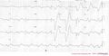
 Wide complex tachycardia: ventricular or supraventricular? ... Case provided by Kees-Jan Royaards, MD
|
 Myocardial infarction or not? ... Case provided by Kees-Jan Royaards, MD
|
 Conduction trouble ... Case provided by Kees-Jan Royaards, MD
|
 Sinus rhythm or not? ... Case provided by JSSG de Jong, MD
|
 A patient presented with a broad complex tachycardia... Case provided by
|
Electrolyte Disorders
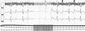
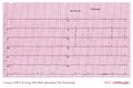
 Try to interprete this ECG using the 7+2 step method Look at the consecutive ECGs in this patient. Case provided by C.J. Royaards, MD
|
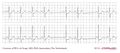
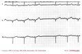
 Look at the consecutive ECGs in this patient. What electrolyte disturbance do you expect? Case provided by C.J. Royaards, MD
|
Miscellaneous
Click on the text below the ECG for the answers. Click on the ECG for enlargement of the ECG itself...
 Case provided by
|
 Typical Brugada syndrome ST segments in right precordial ECG leads (on spot diagnosis) aka 'type-1 Brugada ECG' with 1st degree AV block and broad P-waves. Case provided by W.G. de Voogt, MD, PhD
|
 A regular small-QRS tachycardia at about 300bpm with normal looking QRS complexes is most likely an Atrial flutter with 1:1 conduction over the AV node. Case provided by W.G. de Voogt, MD, PhD
|
Rhythm Puzzles by Prof. A.A.M. Wilde
 A 95-year-old woman presents with palpitations. She has no relevant medical history and the present complaint is several days old... Case provided by A.A.M. Wilde
|
 A 71-year-old female patient presents to your outpatients clinic with an irregular heart rhythm. The complaints started a few weeks ago and seem to worsen... Case provided by A.A.M. Wilde
|
 An 83-old-female patient presents to your outpatient clinic with a history of syncope. It has occurred three times in the last few months and there were no specific triggers... Case provided by A.A.M. Wilde
|
 A 57-year-old man collapsed after one hour of angina symptoms in the presence of the alarmed ambulance personnel. He had never had any complaints... Case provided by N.J.W. Verouden, R.J. de Winter, A.A.M. Wilde
|
 A 44-year-old male presented with palpitations. His medical history was otherwise uneventful and the family history revealed pacemaker... Case provided by A.A.M. Wilde, J. Hrudova, J.G.M. Tans
|
 A 73-year-old man presents with palpitations (irregular heartbeat). There is no medical history. Physical examination reveals no abnormalities... Case provided by A.A.M. Wilde
|
 A 55-year-old male without cardiac history is complaining of an irregular heartbeat. Physical examination and an echo reveal no abnormalities... Case provided by A.A.M. Wilde
|
 A 36-year-old male comes to your outpatient clinic because of his family history. His father died suddenly at age 43 and his brother died suddenly at age 39... Case provided by A.A.M. Wilde
|
 A 38-year-old male patient presents with palpitations. He is not suffering from syncope or dizziness and has no other complaints... Case provided by A.A.M. Wilde
|
 A 75-year-old lady presented with palpitations. Her medical history reveals cardiac surgery with a mitral valve repair combined... Case provided by A.A.M. Wilde, H.H.D. Idzerda
|
 A boy with a birth weight of 3.030 g was born by caesarean section at 33 weeks of gestation because of bradycardia and severe... Case provided by A.A.M. Wilde, N.A. Blom
|
 An 86-year-old man presents in your outpatient clinic with stable angina pectoris (NYHA 2/4). There is no further medical history... Case provided by A.A.M. Wilde
|
 A 14-year-old girl complained of palpitations that occurred numerous times a week. There were no special triggers and, usually... Case provided by A.A.M. Wilde, M. Cuppen, J.L.R.M. Smeets
|
 A 43-year-old man came to the outpatient clinic for preventive cardiovascular (CV) screening. During the last five years he... Case provided by N.M. Panhuyzen-Goedkoop, L.R.C. Dekker, A.A.M. Wilde
|
 A 47-year-old lady presents with an ‘abnormal ECG’. In her family three daughters suffer from a long-QT syndrome type 2... Case provided by A.A.M. Wilde
|
 A 63-year-old female was referred to our outpatient clinic with symptoms of palpitations. These had been occurring in paroxysms... Case provided by T.A. Simmers, A.A.M. Wilde
|
 A 34-year old man comes to your office. He has read in a newspaper about the familial occurrence of sudden death. He suffered a... Case provided by A.A.M. Wilde, H.L. Tan
|
 A 28-year-old male was referred because of an abnormal ECG. He was having occasional palpitations. His family history is unremarkable... Case provided by A.A.M. Wilde, L.R.C. Dekker
|
 A 90-year-old lady presented with chest pain which had a sudden onset in the middle of the night. There were no other symptoms... Case provided by A.A.M. Wilde
|
 A 65-year-old male presented with palpitations and dizziness of sudden onset, without any associated chest pain or other symptoms... Case provided by A.A.M. Wilde
|
 A 54-year-old man presented with palpitations. He had no other symptoms. Physical examination revealed, with the exception of a... Case provided by A.A.M. Wilde, R.H. Bakker
|
 A 63-year-old lady presented with episodic chest pain without specific triggers (in particular no relation with exercise)... Case provided by A.A.M. Wilde, R.H.J. Peters
|
 An otherwise healthy 57-year-old lady presented with palpitations without dizziness. The symptoms had been present for a couple... Case provided by A.A.M. Wilde, L.R.C. Dekker
|
 A 28-year-old, wheelchair bound, female patient with Friedreich’s ataxia presents with an irregular heartbeat... Case provided by A.A.M. Wilde
|
 A 61-year-old male was referred with symtoms of exertional dyspnoea and palpitations. He had suffered an anterior myocardial... Case provided by T.A. Simmers
|
 A 64-year-old man had an inferior myocardial infarction ten years ago. Lately he has been having palpitations with occasional dizziness... Case provided by A.A.M. Wilde, R.B.A. van den Brink
|
 An otherwise healthy 32-year-old male was referred with palpitations. Attacks had been occurring monthly for several years... Case provided by A.A.M. Wilde, R.B.A. van den Brink
|
 An otherwise healthy 66-year-old male was referred with complaints of central chest pain. He was not on any medication... Case provided by T.A. Simmers, A.A.M. Wilde
|
 A 20-year-old male is having palpitations. They occur without a specific trigger, although episodes are sometimes related to... Case provided by A.A.M. Wilde, R.B.A. van den Brink
|
 An otherwise healthy 24-year-old man was referred to our hospital because of drug refractory spells of palpitations accompanied by dizziness... Case provided by A.A.M. Wilde, R.B.A. van den Brink
|
 A 33-year-old lady visited the cardiologist because of the sudden death of her brother at age 35. He died while watching TV... Case provided by A.A.M. Wilde, Y.M. Pinto
|
 A 46-year-old male was admitted to our emergency room with dyspnoea. His medical history included congestive heart failure with a left ventricular ejection Case provided by T.A. Simmers, A.A.M. Wilde
|
 A 30-year-old woman presents with repeated syncope. Her symptoms started a few months ago without a particular trigger... Case provided by A.A.M. Wilde and H. Tan
|
 A 65-year-old woman was admitted because of recurrent syncope. Her complaints were difficult to interpret due to mental... Case provided by L.R.C. Dekker, R. Tukkie
|
 An 86-year-old man presents in your outpatient clinic with stable angina pectoris (NYHA 2/4). There is no further medical history... Case provided by A.A.M. Wilde
|
 In the setting of family screening, an 84-year-old lady was invited for a cardiogenetic evaluation. Two of her grandchildren... Case provided by A.A.M. Wilde, T.A. Simmers
|
 A 14-year-old boy died suddenly while playing soccer. He was in the middle of a sprint when he suddenly succumbed... Case provided by A.A.M. Wilde, T.A. Simmers
|
 A 27-year-old female was referred to the emergency room with rapid palpitations... Case provided by I.C.D. Westendorp, G.S. de Ruiter, L.V.A. Boersma, E.F.D. Wever
|
 In 2002, an a trial demand inhibited (AAI) pacemaker was implanted in a young male (born 1984) because of a primary arrhythmia disorder. Case provided by R. Tukkie, R. Rienks, A.A.M. Wilde
|
Case reports by W.G. De Voogt, MD, PhD
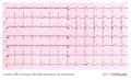
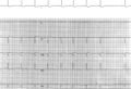
 This Holter registration shows pauses. Case provided by W.G. De Voogt, MD, PhD
|
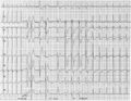
 This ECG shows pauses in the heart rhythm. Case provided by W.G. De Voogt, MD, PhD
|
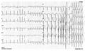
 This tracing shows a pause in the heart rhythm. Case provided by W.G. De Voogt, MD, PhD
|
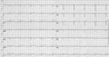
 This is a tracing of a 52 year old male, with viral infection and high fever (40° C), who was admitted to the... Case provided by W.G. De Voogt, MD, PhD
|
Case reports from the ICBA
 Sinus bradycardia, Second degree 2:1 AV block with LBBB in the conducted beats and junctional escape beats with RBBB morphology and fusion beats that mimic normal intraventricular conduction. Case provided by Dr. Alberto Giniger
|
 Sinus rythm, 2:1 AV block with RBBB in the left part of the EKG and after a 1:1 conducted beat with LBBB the 2:1 AV block continues with LBBB. So it is a bilateral branch block. Case provided by Dr. Alberto Giniger
|
 Sinus rythm, high degree AV block, conducted beats with RBBB and ventricular escape beats with LBBB image. Some fusion beats mimic no intraventricular disturbance confundinc as normal conducted beats. Case provided by Dr. Alberto Giniger
|
 Sinus rythm, high degree AV block with unional escape beats and capture beats with RBBB. Case provided by Dr. Alberto Giniger
|
 AV orthodromic tachycardia (WPW syndrome). Case provided by Dr. Alberto Giniger
|
 Ventricular tachycardia or idioventricular rythm during sinus tachycardia with fusion beats. Case provided by Dr. Alberto Giniger
|
 Case provided by Dr. Alberto Giniger
|
 Escape rhythm with left bundle branch block pattern and likely retrograde conducted P waves. Case provided by Dr. Alberto Giniger
|
 Intermittent pre-exitation Case provided by Dr. Alberto Giniger
|
 SA exit block Case provided by Dr. Alberto Giniger
|
 Complete AV block and left ventricle escape. Case provided by Dr. Alberto Giniger
|
 Idionodal escape rythm and ventricular capture by AV nodal reentry... Case provided by Dr. Alberto Giniger
|
 Idioventricular rythm with 1:1 V-A conduction. Case provided by Dr. Alberto Giniger
|
 Mobitz II AV block with LBBB in the conducted beats. Case provided by Dr. Alberto Giniger
|
 Idioventricular accelerated rythm with fusion beats. Case provided by Dr. Alberto Giniger
|
 AV nodal reentry tachycardia. Also look at the below ladder diagram. Case provided by Dr. Alberto Giniger
|
 AV block with idionodal escape (the third P wave is inside the QRS). Case provided by Dr. Alberto Giniger
|
 Sinus bradicardia and isorythmic nodal escapes. Case provided by Dr. Alberto Giniger
|
 Coarse atrial fibrillation mimic atrial flutter. Case provided by Dr. Alberto Giniger
|
 Long QT and VPB secondary to sotalol administration Case provided by Dr. Alberto Giniger
|
 Intermittent WPW Case provided by Dr. Alberto Giniger
|