DV Case 3 Answer
Jump to navigation
Jump to search
| This page is part of De Voogt Archive - Cases
Previous Case: / | Next Case: DVA Case 4 |
Questions
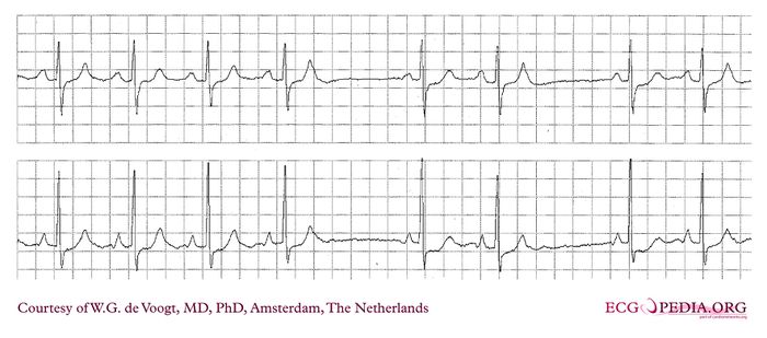
This Holter registration shows pauses.
- Is this SA block?
- Is this AV-block (Wenckebach)
- Is there an other cause for the 2 pauses in this strip?
Answer
- In SA block it is more difficult to appreciate the conduction delay as SA conduction is not presented on a surface ECG. SA block is seen as sudden duplication of the P-P interval, as is the case in 2:1 block, or more difficultly seen as progressive shortening of the P-P interval with a pause thereafter, in case of SA - Wenckebach block. (Definition of a Wenckebach block : decline in progression of the incremental delay).
- Sometimes AV Wenckebach is difficult to see on the ECG as the increment of PR interval can be minimal. The PR interval after a the pause is shorter than in the consecutive beats in the tracing. This could indicate AV Wenckebach. However the conduction in the AV node after a longer pause has recovered more from the preceding depolarization, than during a higher frequency. This makes the short PR interval after a pause is not indicative of AV Wenckebach per se.
- The cause of the interruption in the rhythm is an atrial premature beat (APB), as seen on the T-wave of the last beat before the pause. This T wave is of a different morphology compared to the preceding T-waves, caused by the APB. The correct diagnosis is: blocked atrial premature beats (APB).
This premature beat is not conducted through the AV node, as the AV node is still refractory from the preceding depolarization. The APB however, is conducted retrograde to the SA node, making refractory tissue around the SA-node. This is the cause, the sinus node activity cannot depolarize the atrium and no sinus P-wave is seen.
Note the pause is approximately double the P-P distance preceding it.
