Approach to the Wide Complex Tachycardia
During wide complex tachycardia (heart rate > 100/min, QRS > 0.12 sec) the differentiation between supraventricular and ventricular origin of the arrhythmia is important to guide therapy. Several algorithms have been developed to aid in this differentiation (below). It is important to keep in mind that a good estimate of VT versus SVT can be made based on the clinical vignette:
- 'Horizontal entrance' into the ER. Older patient with previous myocardial infarction = most likely VT
- Younger patient with known paroxysmal tachycardias and who is hemodynamically stable = most like SVT
The ACC algorithm ACC

Brugada criteria

| Morphological criteria (if the above criteria are inconclusive) | ||
|---|---|---|
| LBBB pattern | ||
| Initial R more than 40ms? | Yes => VT |  |
| Slurred or notched downwards leg of S wave in leads V1 or V2 | Yes => VT | |
| Beginning of Q to nadir QS >60 ms in V1 or V2? | Yes => VT | LR >50:1 |
| Q or QS in V6? | Yes => VT | LR >50:1 |
 | ||
| RBBB pattern | ||
| Monofasic R or qR in V1? | Yes => VT | |
| R taller than R' (rabbit-ear sign)? | Yes => VT | LR >50:1 |
| rS in V6? | Yes => VT | LR >50:1 |
 | ||
Ultrasimple Brugada criterion

In 2010 Joseph Brugada et al. published a new criterion to differentiate VT from SVT in wide complex tachycardias: the R wave peak time in Lead II Brugada2. They suggest measuring the duration of onset of the QRS to the first change in polarity (either nadir Q or peak R) in lead II. If the RWPT is ≥ 50ms the likelihood of a VT very high (positive likelihood ratio 34.8). This criterion was successful in their own population of 163 selected patients and is awaiting prospective testing in a larger trial.
Vereckei algorithm Vereckei


Examples
-
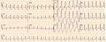 Wide complex tachycardia. No AV dissociation. RBBB. Resembles sinus rhythm from the same patient. Conclusion: SVT with RBBB
 -
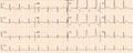 ECG from the same patient in sinus rhythm. The QRS complex is very similiar.
 -
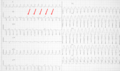 Wide complex tachycardia. LBBB configuration. Absence of RS in the chest leads. AV dissociation is present. Conclusion: VT
 -
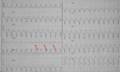 Wide complex tachycardia. LBBB configuration. Absence of RS in the chest leads. AV dissociation is present. Conclusion: VT


Referenties
<biblio>
- ACC pmid=14563598
- Brug1 pmid=2022022
- Vereckei pmid=17272358
- Brugada2 pmid=20215043
</biblio>