Localisation of the origin of a ventricular tachycardia: Difference between revisions
Jump to navigation
Jump to search
mNo edit summary |
mNo edit summary |
||
| (7 intermediate revisions by 2 users not shown) | |||
| Line 1: | Line 1: | ||
[[File:VT_origins.svg|thumb|Areas of the left ventricle where VT's can originate from: The left ventricle is depicted as having been opened. | [[File:VT_origins.svg|thumb|Areas of the left ventricle where VT's can originate from: The left ventricle is depicted as having been opened. Regions are defined as: '''AA''' = antero-apical; '''AB''' = antero-basal; '''AM''' = mid-anterior; '''SA''' = apical septum; '''SB''' = basal-septum; '''SM''' = mid-septum; '''IA''' = inferior apex; '''IB''' = inferior-basal; '''IM''' = mid-inferior. '''Posterior / lateral''' is located on the part between anterior and inferior. Adapted from Miller et al.<cite>Miller</cite>]] | ||
The localisation of the origin (or exit site) of a ventricular tachycardia can be helpful in understanding the cause of the VT and is very helpful when planning an ablation procedure to treat a ventricular tachycardia. | The localisation of the origin (or exit site) of a ventricular tachycardia can be helpful in understanding the cause of the VT and is very helpful when planning an ablation procedure to treat a ventricular tachycardia. | ||
Using this approach and the | The steps to finding the exit site are: | ||
[[File:LBBB_VT_i.svg|thumb|left|300px|Localising the VT exit in LBBB VT with negative QRS complexes inferior]] | # What is the bundle branch block (BBB) configuration? | ||
[[File:LBBB_VT_p.svg|thumb|none|300px|Localising the VT exit in LBBB VT with positive QRS complexes inferior]] | # What is the inferior lead QRS complex polarity? | ||
[[File:RBBB_VT.svg|thumb|300px|left|Localising the VT exit in RBBB VT with | # What is the lead I QRS complex polarity? | ||
[[File:RBBB_VT2.svg|thumb|300px|none|Localising the VT exit in RBBB VT with | # What is the lead aVL QRS complex polarity? | ||
# What is the lead aVR QRS complex polarity? | |||
# Where is the R-wave transition point? | |||
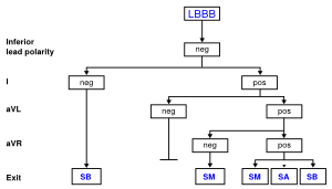
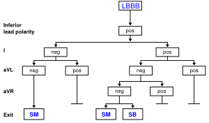
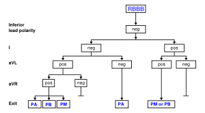
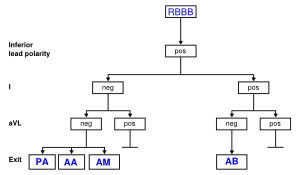
Using this approach and the algorithms below <cite>segal</cite> the exit site can be estimated with reasonable accuracy (PPV around 70%). In these algorhythms, bundle branch block was defined as “left” or “right” based on QRS morphology in lead V1; right bundle branch block (RBBB) pattern was defined by a mono-, bi-, or triphasic R wave or qR in V1; LBBB pattern was defined by a QS, rS, or qrS in V1. | |||
[[File:LBBB_VT_i.svg|thumb|left|300px|Localising the VT exit in LBBB VT with negative QRS complexes inferior. Adapted from Segal et al.<cite>segal</cite>]] | |||
[[File:LBBB_VT_p.svg|thumb|none|300px|Localising the VT exit in LBBB VT with positive QRS complexes inferior. Adapted from Segal et al.<cite>segal</cite>]] | |||
[[File:RBBB_VT.svg|thumb|300px|left|Localising the VT exit in RBBB VT with negative QRS complexes inferior. Adapted from Segal et al.<cite>segal</cite>]] | |||
[[File:RBBB_VT2.svg|thumb|300px|none|Localising the VT exit in RBBB VT with positive QRS complexes inferior. Adapted from Segal et al.<cite>segal</cite>]] | |||
==References== | ==References== | ||
<biblio> | <biblio> | ||
Latest revision as of 15:44, 8 March 2023

The localisation of the origin (or exit site) of a ventricular tachycardia can be helpful in understanding the cause of the VT and is very helpful when planning an ablation procedure to treat a ventricular tachycardia.
The steps to finding the exit site are:
- What is the bundle branch block (BBB) configuration?
- What is the inferior lead QRS complex polarity?
- What is the lead I QRS complex polarity?
- What is the lead aVL QRS complex polarity?
- What is the lead aVR QRS complex polarity?
- Where is the R-wave transition point?
Using this approach and the algorithms below segal the exit site can be estimated with reasonable accuracy (PPV around 70%). In these algorhythms, bundle branch block was defined as “left” or “right” based on QRS morphology in lead V1; right bundle branch block (RBBB) pattern was defined by a mono-, bi-, or triphasic R wave or qR in V1; LBBB pattern was defined by a QS, rS, or qrS in V1.


References
<biblio>
- segal pmid=17338765
- Miller pmid=3349580
</biblio>