Intraventricular Conduction: Difference between revisions
| Line 59: | Line 59: | ||
{{clr}} | {{clr}} | ||
=== | ===LPFB=== | ||
Criteria | ;Criteria for posterior fascicular block: | ||
:right [[heart axis|axis devation]] >+120°; | |||
:deep S in I; | |||
:small q in III; | |||
:no or very few QRS widening; | |||
:Right ventricular hypertrophy and previous lateral myocardial infarction have been excluded | |||
==References== | ==References== | ||
Revision as of 17:45, 20 May 2007
| Author(s) | J.S.S.G. de Jong, MD | |
| Moderator | J.S.S.G. de Jong, MD | |
| Supervisor | ||
| some notes about authorship | ||
Conduction delay
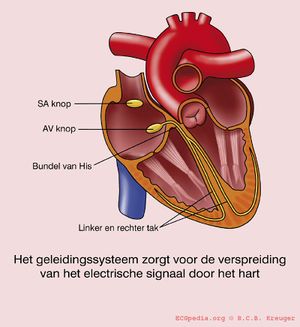
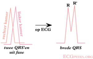
If the QRS complex is wider than 0.12 seconds this is mostly caused by a delay in the conduction tissue of one of the bundle branches:
- Left Bundle Branch Block (LBBB))
- Right Bundle Branch Block(RBBB)
- Interventricular conduction delay
A right or left axis rotation can be caused by a:
Sometimes this conduction delay is frequency-dependent : the bundle branch block occurs only at higher heart rates and disappears at slower heart rates.
LBBB vs RBBB
Check V1 for QRS > 0,12 sec.
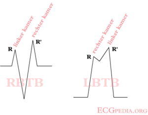
When the last QRS in V1 is below the baseline (moving away from V1), a LBBB is the most likely diagnosis.
When the last activity is above the baseline, it's a RBBB.
If the QRS > 0.12 sec. but the morphological criteria of LBBB or RBBB do not apply, it is called 'interventriculair conduction delay', a general term.
LBBB
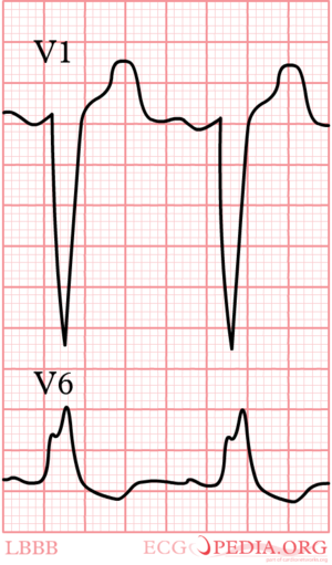
- Criteria for left bundle branch block (LBBB) [1]
- QRS >0,12 sec
- Broad monomorphic R waves in I and V6 with no Q waves
- Broad monomorphic S waves in V1, may have a small r wave
In left bundle branch block (LBBB) the conduction in the left bundle is slow. This results in delayed depolarisation of the left ventricle, especially the left lateral wall. The electrical activity in the left lateral wall is unopposed by the usual right ventricular electrical activity. The last activity on the ECG thus goes to the left or away from V1. Once you remember this, LBBB is easy to understand.
RBBB
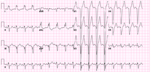
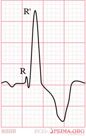
- Criteria for right bundle branch block (RBBB) [1]
- QRS >0,12 sec
- Slurred S wave in lead I and V6
- RSR'-pattern in V1 where R' > R
Again, watch V1. In right bundle branch block (RBBB) the conduction in the bundle to the right ventricle is slow. As the right ventricles depolarizes, the left ventricle is often halfway finished and few counteracting electrical activity is left. The last electrical activity is thus to the right, or towards lead V1. In RBBB the QRS complex in V1 is allways markedly positive.
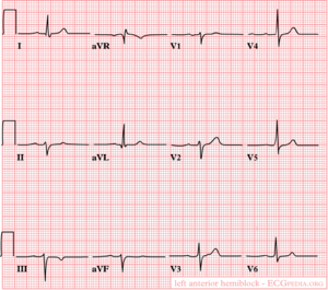
LAFB
- Criteria for left anterior fascicular block
- left axis deviation (<-30°)
- no or very small S in lead I
- normal small q in lead I
- S > R in leads II and III
- no or very few QRS widening
In left anterior fascicular block the anterior part (fascicle) of the left bundle is slow. This results in delayed depolarisation of the upper anterior part of the left ventricle. On the ECG this results in left axis deviation. The QRS width is <0,12 seconds in isolated LAFB.
LPFB
- Criteria for posterior fascicular block
- right axis devation >+120°;
- deep S in I;
- small q in III;
- no or very few QRS widening;
- Right ventricular hypertrophy and previous lateral myocardial infarction have been excluded
