Localisation of the origin of a ventricular tachycardia: Difference between revisions
Jump to navigation
Jump to search
mNo edit summary |
mNo edit summary |
||
| Line 5: | Line 5: | ||
Using this approach and the algorithms below <cite>segal</cite> the exit site can be estimated with reasonable accuracy (PPV around 70%). In these algorhythms, bundle branch block was defined as “left” or “right” based on QRS morphology in lead V1; right bundle branch block (RBBB) pattern was defined by a mono-, bi-, or triphasic R wave or qR in V1; LBBB pattern was defined by a QS, rS, or qrS in V1. | Using this approach and the algorithms below <cite>segal</cite> the exit site can be estimated with reasonable accuracy (PPV around 70%). In these algorhythms, bundle branch block was defined as “left” or “right” based on QRS morphology in lead V1; right bundle branch block (RBBB) pattern was defined by a mono-, bi-, or triphasic R wave or qR in V1; LBBB pattern was defined by a QS, rS, or qrS in V1. | ||
[[File:LBBB_VT_i.svg|thumb|left|300px|Localising the VT exit in LBBB VT with negative QRS complexes inferior]] | [[File:LBBB_VT_i.svg|thumb|left|300px|Localising the VT exit in LBBB VT with negative QRS complexes inferior. Adapted from Segal et al.<cite>segal</cite>]] | ||
[[File:LBBB_VT_p.svg|thumb|none|300px|Localising the VT exit in LBBB VT with positive QRS complexes inferior]] | [[File:LBBB_VT_p.svg|thumb|none|300px|Localising the VT exit in LBBB VT with positive QRS complexes inferior. Adapted from Segal et al.<cite>segal</cite>]] | ||
[[File:RBBB_VT.svg|thumb|300px|left|Localising the VT exit in RBBB VT with positive QRS complexes inferior]] | [[File:RBBB_VT.svg|thumb|300px|left|Localising the VT exit in RBBB VT with positive QRS complexes inferior. Adapted from Segal et al.<cite>segal</cite>]] | ||
[[File:RBBB_VT2.svg|thumb|300px|none|Localising the VT exit in RBBB VT with negative QRS complexes inferior]] | [[File:RBBB_VT2.svg|thumb|300px|none|Localising the VT exit in RBBB VT with negative QRS complexes inferior. Adapted from Segal et al.<cite>segal</cite>]] | ||
==References== | ==References== | ||
<biblio> | <biblio> | ||
Revision as of 17:10, 9 May 2010

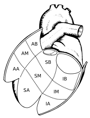
Areas of the left ventricle where VT's can originate from: The left ventricle is depicted as having been opened. Regions are defined as: AA = antero-apical; AB = antero-basal; AM = mid-anterior; SA = apical septum; SB = basal-septum; SM = mid-septum; PA = posterior apex; PB = postero-basal; PM = mid-posterior.. Adapted from Miller et al.[1]
The localisation of the origin (or exit site) of a ventricular tachycardia can be helpful in understanding the cause of the VT and is very helpful when planning an ablation procedure to treat a ventricular tachycardia.
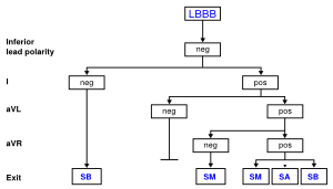
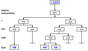
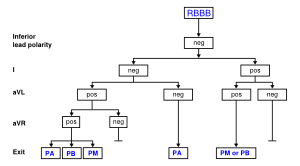
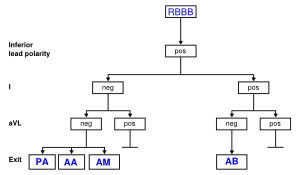
Using this approach and the algorithms below [2] the exit site can be estimated with reasonable accuracy (PPV around 70%). In these algorhythms, bundle branch block was defined as “left” or “right” based on QRS morphology in lead V1; right bundle branch block (RBBB) pattern was defined by a mono-, bi-, or triphasic R wave or qR in V1; LBBB pattern was defined by a QS, rS, or qrS in V1.

Localising the VT exit in LBBB VT with negative QRS complexes inferior. Adapted from Segal et al.[2]

Localising the VT exit in LBBB VT with positive QRS complexes inferior. Adapted from Segal et al.[2]

Localising the VT exit in RBBB VT with positive QRS complexes inferior. Adapted from Segal et al.[2]

Localising the VT exit in RBBB VT with negative QRS complexes inferior. Adapted from Segal et al.[2]
References
- Miller JM, Marchlinski FE, Buxton AE, and Josephson ME. Relationship between the 12-lead electrocardiogram during ventricular tachycardia and endocardial site of origin in patients with coronary artery disease. Circulation. 1988 Apr;77(4):759-66. DOI:10.1161/01.cir.77.4.759 |
- Segal OR, Chow AW, Wong T, Trevisi N, Lowe MD, Davies DW, Della Bella P, Packer DL, and Peters NS. A novel algorithm for determining endocardial VT exit site from 12-lead surface ECG characteristics in human, infarct-related ventricular tachycardia. J Cardiovasc Electrophysiol. 2007 Feb;18(2):161-8. DOI:10.1111/j.1540-8167.2007.00721.x |