Ventricular Tachycardia: Difference between revisions
m (→Examples) |
mNo edit summary |
||
| Line 11: | Line 11: | ||
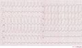
| example = An example of a polymorphic ventricular tachycardia [[Image:vtach.png|250px|Ventricular Tachycardia (VT or V-tach)]] | | example = An example of a polymorphic ventricular tachycardia [[Image:vtach.png|250px|Ventricular Tachycardia (VT or V-tach)]] | ||
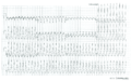
| example2 = Ventricular tachycardia on a 12 lead recording[[Image:12lead_vt1.jpg|thumb|Ventricular tachycardia of 140 bpm with a left bundle branch block and a left axis.]] | | example2 = Ventricular tachycardia on a 12 lead recording[[Image:12lead_vt1.jpg|thumb|Ventricular tachycardia of 140 bpm with a left bundle branch block and a left axis.]] | ||
| animation = < | | animation = <flashow>http://nl.ecgpedia.org/images/7/73/TenTusscherVT.swf|width=300|height=300|quality=best|align=right||</flashow> | ||
| animationdesc = This movie shows a computer model of ventricular tachycardia in the human heart. The VT in this example is initiated by a ventricular extrasystole and maintained by a re-entry rotor inferior in the left ventricle.<cite>tentusscher</cite> In reality, this is possibly not the etiology of VT in diseased hearts, however it is very illustrative. Read [[Copyright|this]] if you want to use this image in a presentation. [[Media:TenTusscherVT.swf|Link to the file / enlargement]] | | animationdesc = This movie shows a computer model of ventricular tachycardia in the human heart. The VT in this example is initiated by a ventricular extrasystole and maintained by a re-entry rotor inferior in the left ventricle.<cite>tentusscher</cite> In reality, this is possibly not the etiology of VT in diseased hearts, however it is very illustrative. Read [[Copyright|this]] if you want to use this image in a presentation. [[Media:TenTusscherVT.swf|Link to the file / enlargement]] | ||
}} | }} | ||
Revision as of 19:17, 1 April 2008
| This is part of: Ventricular Arrhythmias |
| {{{locatieafbeelding}}} | |
| Atrial rate | 60-100 bpm |
| Ventricular rate | 110-250 bpm |
| Regularity | regular |
| Origin | ventricles |
| P-wave | AV-dissociation |
| Effect of adenosine | no rate reduction (can accelerate) |
Example ECG: An example of a polymorphic ventricular tachycardia 
| |
| Example ECG2: Ventricular tachycardia on a 12 lead recording | |

Ventricular tachycardia is defined as a sequence of three or more ventricular beats. The frequency must by higher than 100 bpm, mostly it is 110-250 bpm.
Ventricular tachycardias often origin around old scar tissue in the heart, e.g. after myocardial infarction. Also electrolyte disturbances and ischemia can cause ventricular tachycardias. The cardiac output is often strongly reduced during VT resulting in hypotension and loss of conciousness. VT is a medical emergency as it can deteriorate into Ventricular fibrillation and thus mechanical cardiac arrest.
Ventricular tachycardia can be catechorized as follows:
- Non-sustained VT: three or more ventricular beats with a maximal duration of 30 seconds.
- Sustained VT: a VT of more than 30 seconds duration (or less if treated by electrocardioversion within 30 seconds).
- Monomorphic VT: all ventricular beats have the same configuration.
- Polymorphic VT: the ventricular beats have a changing configuration. The RR interval is 180-600 ms (comparable to a heart rate of 100-333 bpm).
- Biphasic VT: a ventricular tachycardia with a QRS complex that alternates from beat to beat. Associated with digoxin intoxication.
Examples
Ventricular tachycardia of 140 bpm with a left bundle branch block pattern and left heart axis.
Ventricular tachycardia of 250 bpm with a right bundle branch block pattern and right heart axis.
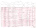
Ventricular tachycardia of 150 bpm with a right bundle branch block pattern and right heart axis. Mind the 5th and 6th complex from the right side. These are fusion complexes. Furthermore this ECG shows baseline drift, which is a technical artefact

