Search results
Jump to navigation
Jump to search
Page title matches
File:2 1 fusion beats.jpg Sinus bradycardia, Second degree 2:1 AV block with LBBB in the conducted beats and junctional escape beats with(1,022 × 684 (135 KB)) - 12:40, 19 February 2010
File:2 1 LBBB RBBB.jpg ...in the left part of the EKG and after a 1:1 conducted beat with LBBB the 2:1 AV block continues with LBBB. So it is a bilateral brunch block.(956 × 548 (81 KB)) - 12:44, 19 February 2010
File:De-Casus1 1 2.jpg (400 × 81 (13 KB)) - 04:33, 10 May 2012


Page text matches
File:2 1 LBBB RBBB.jpg ...in the left part of the EKG and after a 1:1 conducted beat with LBBB the 2:1 AV block continues with LBBB. So it is a bilateral brunch block.(956 × 548 (81 KB)) - 12:44, 19 February 2010
File:ICBA00002.jpg ...in the left part of the EKG and after a 1:1 conducted beat with LBBB the 2:1 AV block continues with LBBB. So it is a bilateral brunch block.(3,000 × 1,797 (1.28 MB)) - 19:20, 23 February 2010
File:ICBA00013.jpg |Description = Idioventricular rythm with 1:1 V-A conduction(3,000 × 1,975 (2.32 MB)) - 10:15, 27 February 2010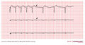
File:E288.jpg 2 to 1 AV block (every other P wave is conducted to the ventricles) 2 to 1 AV block starts after the 5th QRS in this 3 channel recording. The first no 2 to 1 AV block cannot be classified into Mobitz type I or II as we do not know if(3,004 × 1,599 (4.46 MB)) - 07:29, 21 February 2012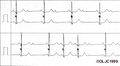
File:E.jpg ...e top tracing and a Mobitz II A/V block on the lower one. Note that with 2:1 block you can not tell if this is a Mobitz I or II. Mobitz II is seen below(470 × 257 (17 KB)) - 06:58, 21 February 2012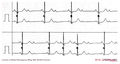
File:E000738.jpg ...e top tracing and a Mobitz II A/V block on the lower one. Note that with 2:1 block you can not tell if this is a Mobitz I or II. Mobitz II is seen below(3,004 × 1,599 (873 KB)) - 03:11, 11 February 2012File:2 1 fusion beats.jpg Sinus bradycardia, Second degree 2:1 AV block with LBBB in the conducted beats and junctional escape beats with(1,022 × 684 (135 KB)) - 12:40, 19 February 2010
File:E00031141.jpg ...in shortness of breath. The electrocardiogram is remarkable as it shows 1:1 retrograde conduction from the ventricle to the atrium, which is best seen(3,220 × 1,815 (5.55 MB)) - 09:53, 17 February 2012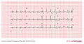
File:E196.jpg ...RS is difficult to determine, but one usually looks at the first 60 ms. (1 1/2 small squares) to determine the axis with a RBBB. If the axis of the firs(3,004 × 1,599 (4.43 MB)) - 00:33, 21 February 2012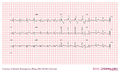
File:E000704.jpg ...RS is difficult to determine, but one usually looks at the first 60 ms. (1 1/2 small squares) to determine the axis with a RBBB. If the axis of the firs(2,644 × 1,599 (3.41 MB)) - 06:12, 10 February 2012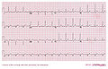
File:DVA2532.jpg |Description = WPW 1(3,000 × 1,922 (2.79 MB)) - 13:22, 8 March 2011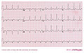
File:DVA2578.jpg |Description = WPW 1(3,000 × 1,930 (2.81 MB)) - 11:57, 9 March 2011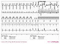
File:DVA2442.jpg |Description = burst 1(3,000 × 2,208 (1.28 MB)) - 14:07, 7 March 2011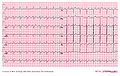
File:DVA2572.jpg |Description = trage flutter 1(3,000 × 1,906 (3.84 MB)) - 11:52, 9 March 2011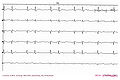
File:DVA2424.jpg |Description = AV wenckebach 1(3,000 × 1,988 (1.87 MB)) - 13:40, 7 March 2011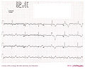
File:DVA2487.jpg |Description = LQT door blok 1(3,000 × 2,451 (1.28 MB)) - 11:53, 8 March 2011
File:DVA2508.jpg |Description = SSS met AV gel st 1(3,000 × 2,571 (4.01 MB)) - 12:33, 8 March 2011
File:DVA2541.jpg |Description = Bifasc block met 2 op 1(3,000 × 1,930 (3.73 MB)) - 10:55, 9 March 2011
File:DVA2595.jpg |Description = AF en PM bij HCM 1(3,000 × 1,925 (3.51 MB)) - 12:21, 9 March 2011
File:DVA2485.jpg |Description = HR B 76 YRS after 1 hour(3,000 × 1,913 (2.65 MB)) - 11:52, 8 March 2011


