Palpitations Again, Have a Closer Look: Difference between revisions

From ECGpedia
Jump to navigation Jump to search
mNo edit summary
 
(One intermediate revision by the same user not shown)
Line 19: Line 19:


[[Puzzle_2006_11_393_Answer|Answer]]
[[Puzzle_2006_11_393_Answer|Answer]]
[[Image:Puzzle_2006_11_393_fig2.jpg|Figure 3. Detail from the aVR, aVL and aVF leads during the tachycardia. The arrows point to the (retrograde) P top.|thumb]]
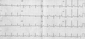
Figure 1 shows a narrow-complex tachycardia with an
RR interval of 390 msec (rate 155 beats/min). In the
latter part the arrhythmia is slightly slower (RR interval
410). In the differential diagnosis of any narrowcomplex
tachycardia, the position of the P wave (and
its number) is critical for the correct diagnosis.
A close look for the P waves reveals a P wave in
every other T wave in the left part of the ECG. After
the 15th QRS complex, the P wave is present in every
T wave (see rhythm strip lead II at the bottom of the
ECG and figure 3, arrows). Comparison of the T waves
with and without superimposed P waves reveals a
negative morphology of the P wave in lead II. In lead
aVR the morphology is clearly positive. The timing of
the P wave is between 40 and 80 ms after the QRS
complex.
The number of P waves indicates a dissociation
between the atrial and ventricular rate and the morphology
indicates retrograde (VA) conduction. The
presence of VA dissociation is only compatible with
AV-nodal reentrant tachycardia and it excludes AVreentry
tachycardia (orthodromic circus movement
tachycardia), atrial tachycardia and atrial flutter, the
most important differential diagnostic entities for a
narrow-complex tachycardia with the present rate. A
rare alternative diagnosis would be a His-bundle tachycardia
with 2:1 VA conduction. Actually the position
of the P wave would be in favour of this diagnosis.
An invasive electrophysiological study revealed the
presence of dual nodal characteristics and, under
isoprenaline, the induction of an AV-nodal reentrant
tachycardia. Slow pathway ablation successfully
modified the arrhythmogenic substrate.

Latest revision as of 20:05, 25 January 2010

Author(s) A.A.M. Wilde, L.R.C. Dekker
NHJ edition: 2006:11,393
These Rhythm Puzzles have been published in the Netherlands Heart Journal and are reproduced here under the prevailing creative commons license with permission from the publisher, Bohn Stafleu Van Loghum.
The ECG can be enlarged twice by clicking on the image and it's first enlargement
Figure 1. The ECG on admission
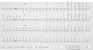
Figure 2. The ECG shortly thereafter

An otherwise healthy 57-year-old lady presented with palpitations without dizziness. The symptoms had been present for a couple of years but the number of episodes had increased recently. Onset and termination were always sudden without specific triggers. Physical examination on admission revealed no specific abnormalities except for a rapid pulse (150 beats/min). Blood pressure was normal. The ECG is shown in figure 1 and a few minutes later figure 2 was recorded without any specific intervention.

What is your diagnosis?
Answer