Atrial Premature Complexes
| This is part of: Supraventricular Rhythms |


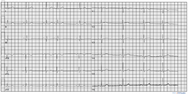
Premature atrial complexes origin from an ectopic pacing region in the atria. They are an example of ectopic beats. The result is a p-wave with often a different morphology from the preceding ones.
If a premature atrial complex follows early after a previous sinus beat some of the the conducting tissues below may not conduct. A premature atral complex can therefore have different fates (see figure):
- Normally conducted
- Conducted with aberrancy. Mostly right bundle branch block aberrancy as the RBBB has a longer refractory period.
- Non-conducted. If the premature beat is very early, the AV node is refractory (cannot conduct) and the beat is not followed by a QRS complex. A non-conducted premature atrial beat is often confused with type II second degree AV block where a normal sinus beat is not followed by a QRS complex.
A premature atrial complex is usually followed by a noncompensatory pause caused by the fact that atrial depolarization enters the sinus node and resets the sinus rhythm.
Premature atrial complexes are common and usually benign. Frequent atrial ectopic beats (>100 / 24hrs) in a population of patients presented to a cardiovascular hospital wass associated with a small chance of 1% of developing atrial fibrillation in the following year AES
See also:
References
<biblio>
- AES pmid=23499279
</biblio>