McGill Case 98: Difference between revisions
Jump to navigation
Jump to search
(Created page with "{{McGillcase| |previouspage= McGill Case 97 |previousname= McGill Case 97 |nextpage= McGill Case 99 |nextname= McGill Case 99 }} [[File:E000798.jpg|thumb|600px|left|This card...") |
No edit summary |
||
| Line 6: | Line 6: | ||
}} | }} | ||
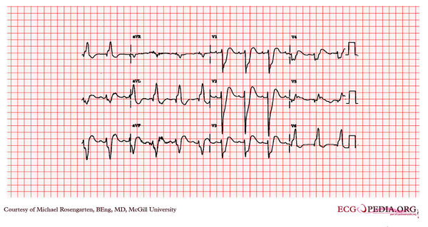
[[File: | [[File:E000799.jpg|thumb|600px|left|This cardiogram shows normal VVI pacemaker function. The patient's native EKG seems relatively normal. Of interest notice the "capture beats" where the narrow complexes that are conducted through the A/V node appear earlier than the paced beats but are narrow. This suggests that there is no retrograde conduction from the ventricle to the atrium as the sinus node seems unaffected by the ventricular pacing. This is some what similar to the case of patients with VT where capture beats are seen (very rarely).]] | ||
Revision as of 01:25, 15 February 2012
|

This cardiogram shows normal VVI pacemaker function. The patient's native EKG seems relatively normal. Of interest notice the "capture beats" where the narrow complexes that are conducted through the A/V node appear earlier than the paced beats but are narrow. This suggests that there is no retrograde conduction from the ventricle to the atrium as the sinus node seems unaffected by the ventricular pacing. This is some what similar to the case of patients with VT where capture beats are seen (very rarely).