McGill Case 86: Difference between revisions
Jump to navigation
Jump to search
(Created page with "{{McGillcase| |previouspage= McGill Case 85 |previousname= McGill Case 85 |nextpage= McGill Case 87 |nextname= McGill Case 87 }} [[File:E000786.jpg|thumb|600px|left|This rhyt...") |
No edit summary |
||
| Line 6: | Line 6: | ||
}} | }} | ||
[[File: | [[File:E0007871.jpg|thumb|600px|left|This rhythm strip shows sinus rhythm with salvos of PVCS. Note that you can track the P wave activity in the ventricular complexes and that there is a ventricular capture beat (6th beat). This capture beat confirms that the wide beats are ventricular and also that there is probably no retrograde A/V conduction.]] | ||
[[File:E0007872.jpg|thumb|600px|left|This rhythm strip shows sinus rhythm with salvos of PVCS. Note that you can track the P wave activity in the ventricular complexes and that there is a ventricular capture beat (6th beat). This capture beat confirms that the wide beats are ventricular and also that there is probably no retrograde A/V conduction.]] | |||
Revision as of 03:01, 15 February 2012
|

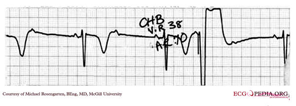
This rhythm strip shows sinus rhythm with salvos of PVCS. Note that you can track the P wave activity in the ventricular complexes and that there is a ventricular capture beat (6th beat). This capture beat confirms that the wide beats are ventricular and also that there is probably no retrograde A/V conduction.

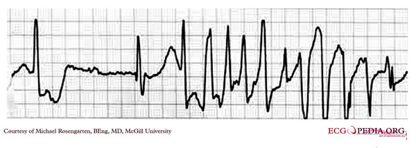
This rhythm strip shows sinus rhythm with salvos of PVCS. Note that you can track the P wave activity in the ventricular complexes and that there is a ventricular capture beat (6th beat). This capture beat confirms that the wide beats are ventricular and also that there is probably no retrograde A/V conduction.