McGill Case 35: Difference between revisions
Jump to navigation
Jump to search
No edit summary |
No edit summary |
||
| Line 6: | Line 6: | ||
}} | }} | ||
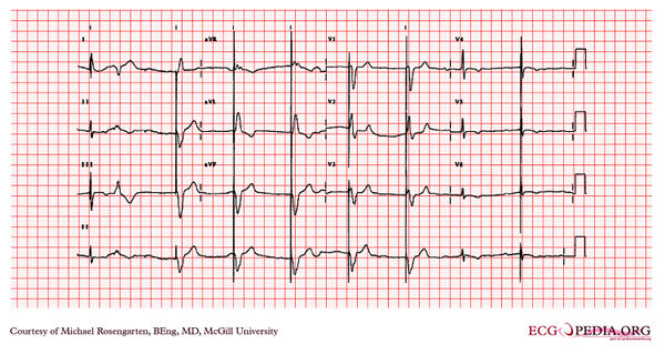
[[File: | [[File:E000735.jpg|thumb|600px|left|This is an electrocardiogram from a 87 year old man with a history of atrial fibrillation. His medications were coumadin and Monopril. | ||
The cardiogram shows sinus rhythm with rate of about 50/min, and a marked first degree heart block with a pr interval of about 350ms. | |||
The first complex on the left is a fusion between the patient's native QRS and the pacemaker spike (this is nomal operation) this is followed by a PVC. Note the small blip following the PVC is artifact and is not a failure to capture of the pacemaker. The pacemaker is working well as a VVI pacer set at 50/min. The large spikes suggest a unipolar lead.]] | |||
Latest revision as of 05:33, 10 February 2012
|

This is an electrocardiogram from a 87 year old man with a history of atrial fibrillation. His medications were coumadin and Monopril. The cardiogram shows sinus rhythm with rate of about 50/min, and a marked first degree heart block with a pr interval of about 350ms. The first complex on the left is a fusion between the patient's native QRS and the pacemaker spike (this is nomal operation) this is followed by a PVC. Note the small blip following the PVC is artifact and is not a failure to capture of the pacemaker. The pacemaker is working well as a VVI pacer set at 50/min. The large spikes suggest a unipolar lead.