File:E338.jpg
Original file (3,004 × 1,599 pixels, file size: 4.67 MB, MIME type: image/jpeg)
Summary
| Description |
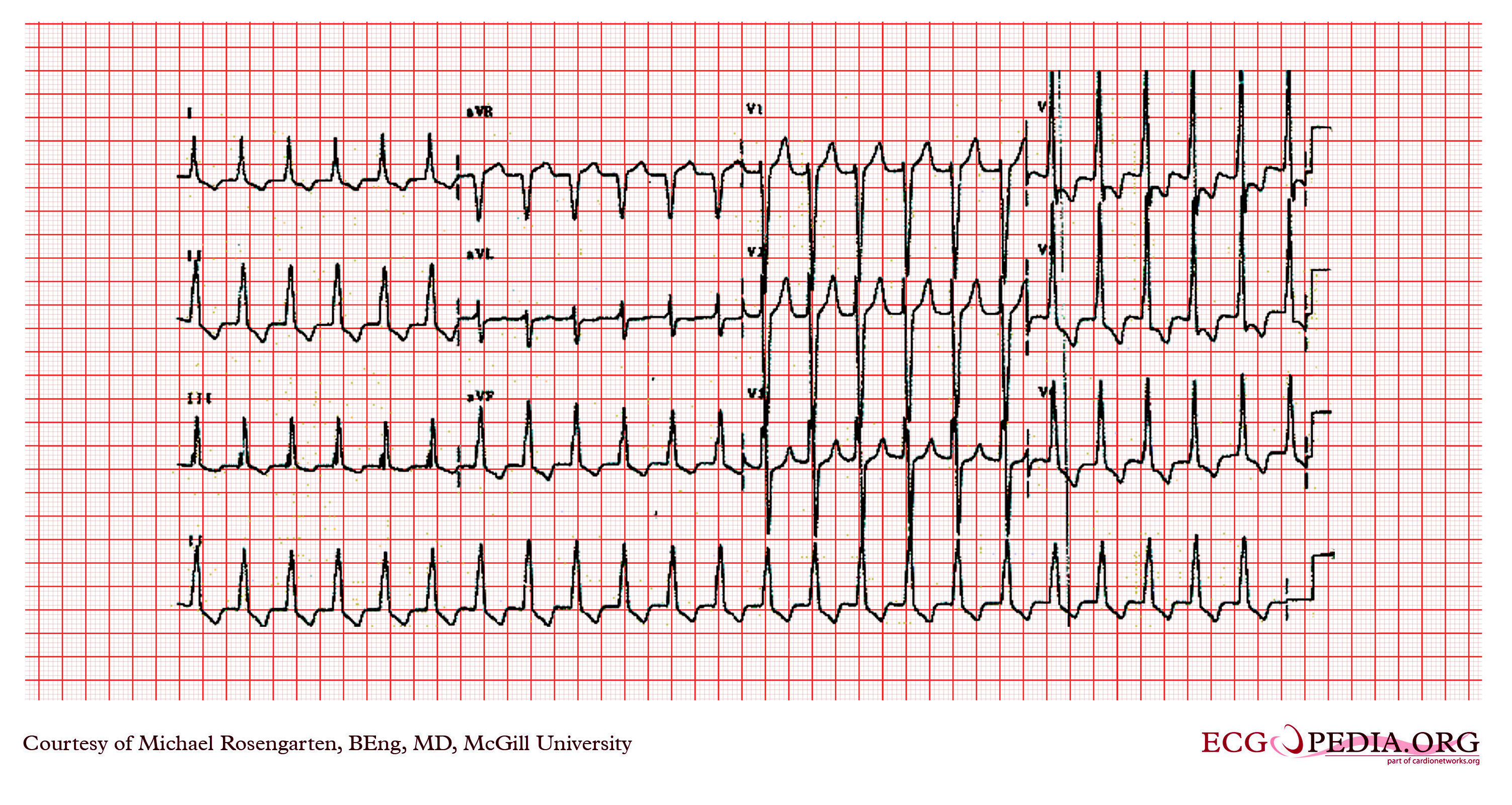
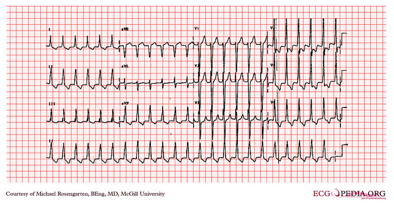
This is an electrocardiogram from an 82 year old man with a history of lung disease and renal failure. At the time of this recording the patient was on Iron, Lasix, and bronchodilators. The EKG shows a regular rhythm at a rate of 141/min. This patient had been in atrial fibrillation in the past and the rhythm here is probably atrial flutter with 2:1 block although no flutter waves are seen. The QRS duration is widened at 105 ms and the tall R waves in V5 and S waves in V1 and V2 and the ST depression in the absence of digoxin suggest left ventricular hypertrophy. The QRS is too narrow for ventricular tachycardia and the brief R wave in V2 with the clean down-stroke of the V2 S wave argue against a ventricular origin. The patient was placed on digoxin with a slowing of the ventricular rate and a rhythm that was clearly atrial fibrillation and with a QRS with the same morphology. |
|---|---|
| Category | |
| Source |
EKG World Encyclopedia http://cme.med.mcgill.ca/php/index.php , courtesy of Michael Rosengarten BEng, MD.McGill |
| Date |
2012 |
| Author |
Michael Rosengarten BEng, MD.McGill |
| Permission |
Creative Commons Attribution Noncommercial Share-Alike License |
File history
Click on a date/time to view the file as it appeared at that time.
| Date/Time | Thumbnail | Dimensions | User | Comment | |
|---|---|---|---|---|---|
| current | 10:42, 21 February 2012 | | 3,004 × 1,599 (4.67 MB) | DarrelC (talk | contribs) |
You cannot overwrite this file.
File usage
The following page uses this file: