File:E264.jpg
Original file (3,004 × 1,599 pixels, file size: 4.42 MB, MIME type: image/jpeg)
Summary
| Description |
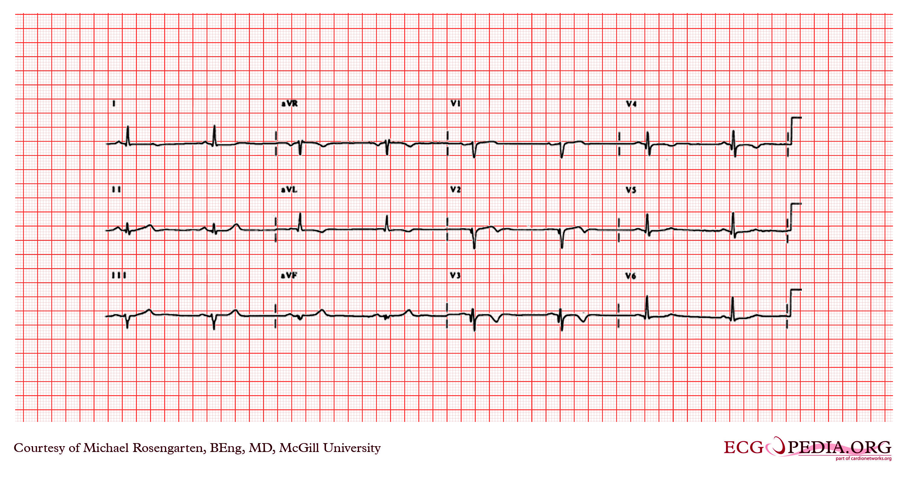
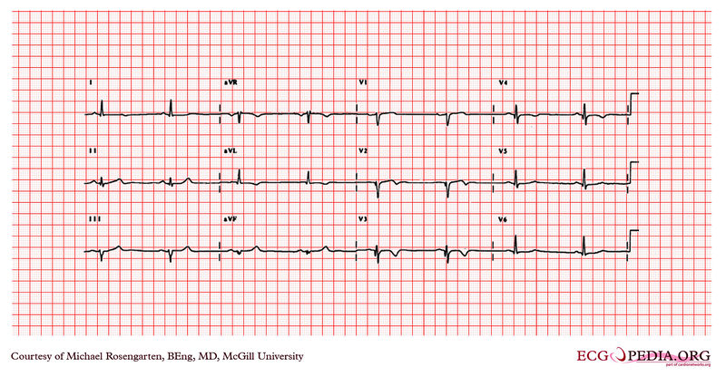
The cardiogram shows sinus bradycardia at 47/min. and a poor r wave progression in the anterior chest leads with Q waves in leads V2 to V4 which are diagnostic of anterior myocardial infarction. Note that unlike the normal septal Q waves that start later in the progression of the chest leads and at the same time grow larger, the Q waves in this patient are abnormal because they are present in leads V2, V3, and V4 and are larger than those in V5 and V6. The cardiogream also shows abnormal T wave inversion and slicht ST ellivation in leads V1 to V3. |
|---|---|
| Category | |
| Source |
EKG World Encyclopedia http://cme.med.mcgill.ca/php/index.php , courtesy of Michael Rosengarten BEng, MD.McGill |
| Date |
2012 |
| Author |
Michael Rosengarten BEng, MD.McGill |
| Permission |
Creative Commons Attribution Noncommercial Share-Alike License |
File history
Click on a date/time to view the file as it appeared at that time.
| Date/Time | Thumbnail | Dimensions | User | Comment | |
|---|---|---|---|---|---|
| current | 06:42, 21 February 2012 | | 3,004 × 1,599 (4.42 MB) | DarrelC (talk | contribs) |
You cannot overwrite this file.
File usage
The following page uses this file: