File:Aivr.jpg: Difference between revisions
Jump to navigation
Jump to search
m (New page: ==Description== An example of accelerated idioventricular rhythm in a patient who was treated with primary PCI after anterior myocardial infarction due to a proximal LAD le...) |
(No difference)
|
Latest revision as of 07:57, 26 July 2007
Description
An example of accelerated idioventricular rhythm in a patient who was treated with primary PCI after anterior myocardial infarction due to a proximal LAD lesion. The first 5 beats and last 9 beats are AIVR. In between two narrow beats are seen of which the second beat is probably a normal sinus beat. AV Dissociation can be seen in leads V1 and V2.
File history
Click on a date/time to view the file as it appeared at that time.
| Date/Time | Thumbnail | Dimensions | User | Comment | |
|---|---|---|---|---|---|
| current | 12:30, 10 April 2010 | 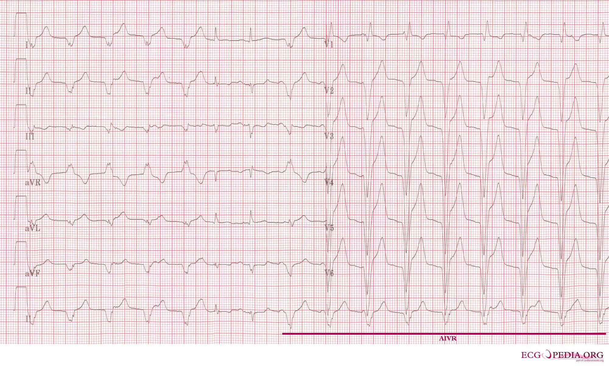 | 2,000 × 1,204 (554 KB) | (username removed) |
File usage
The following page uses this file: