File:E0003143.jpg

Original file (3,004 × 1,599 pixels, file size: 4.42 MB, MIME type: image/jpeg)
Summary
| Description |
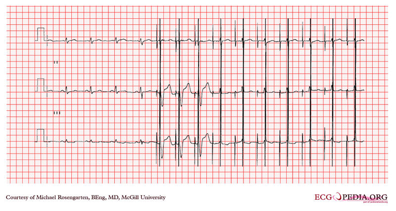
This is a rhythm strip from a patient with a newly implanted ventricular lead. The patient also has an atrial lead. His pacemaker is a dual chamber DDD Medtronic pacemaker. A magnet was placed over the pacemaker in the middle of the recording. This recording shows an important aspect of magnet testing. First the magnet turns off the sensing circuit and makes the pacemaker pace in a DOO mode. Second there are first 3 A/V paced complexes at 100/min. This identifies the beginning of the magnet application, and also does a crude check on threshold by reducing the pulse width by 25% on the 3rd set of spikes. Most important though is that the A/V interval has been shortened to ensure that ventricular capture is seen. Not that in the following complexes the conduction of paced "p" wave captures the ventricle and makes it impossible to determine if there is capture. This is critical in this case both to ensure capture and perhaps more important, to ensure that the paced complex has a left bundle branch morphology confirming that the lead is in the right sided chambers. |
|---|---|
| Category | |
| Source |
EKG World Encyclopedia http://cme.med.mcgill.ca/php/index.php , courtesy of Michael Rosengarten BEng, MD.McGill |
| Date |
2012 |
| Author |
Michael Rosengarten BEng, MD.McGill |
| Permission |
Creative Commons Attribution Noncommercial Share-Alike License |
File history
Click on a date/time to view the file as it appeared at that time.
| Date/Time | Thumbnail | Dimensions | User | Comment | |
|---|---|---|---|---|---|
| current | 22:44, 18 February 2012 |  | 3,004 × 1,599 (4.42 MB) | DarrelC (talk | contribs) |
You cannot overwrite this file.
File usage
The following page uses this file: