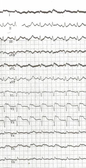
Answers example 1 question 1
Jump to navigation
Jump to search
Answers and below the case will continue!
- Describe the ECG according the 7 + 2 step plan
- Rhythm
- This is a regular rhythm and every QRS complex has a P-wave in front of it. The P wave us positive in II, III and AVF and comes from the sinusnode. So it's a sinusrhythm.
- Heartrate.
- Use the 'counting methode' (3 large grids ~> 300-150-100), so 100/min.
- Conductiontimes (PQ,QRS,QT)
- PQ-time=0.16sec (4 small grids), QRS duration=0.10sec, QT time=280ms, QTc interval=361 ms
- Heart axis
- Isoelectric in I, positive in II, III and AVF. Therefore it is a vertical heartaxis.
- P wave morphology
- The p wave is possibly > 2,5 mm in II (hard to see, a good millimetergrid is lacking), so there could be right atrium overload.
- QRS morphology
- Pathologic Q in AVL, V1-V3 and possibly V4-5 (poor quality). Hardly any precordial R-wave progression.
- ST morphology
- ST elevation in V2-V5 and in I,AVL.
- No prior ECG to compare
- Conclusion. What's going on?
- Rhythm
Answer: A large anterior wall infraction
Additions: and possibly right atriumoverload caused by backwardfailure of the left ventricle.
