Answer MI 8

From ECGpedia
Revision as of 09:26, 11 November 2008 by Drj (talk | contribs) (New page: {{Case| |previouspage= MI 7 |previousname= MI 7 |nextpage=MI 9 |nextname=MI 9 }} '''Where is this myocardial infarction located?''' [[Image:ami0008.jpg|700px|thumb|left|ECG MI 8. Click on...)
(diff) ← Older revision | Latest revision (diff) | Newer revision → (diff)
Jump to navigation Jump to search
This page is part of Cases and Examples

Previous ECG: MI 7 | Next ECG: MI 9

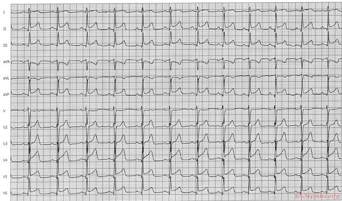
Where is this myocardial infarction located?

ECG MI 8. Click on image for enlargement.


Answer

Culprit lesion: LAD

  1. sinus rhythm
  2. about 75/min
  3. normal conduction
  4. intermediate axis
  5. normal p wave morphology
  6. No pathologic Q or LVH.
  7. ST elevation in II, III, AVF and V4-V6. ST depression in AVR. The ST vector is pointing downwards.
  • Conclusion: Distal LAD occlusion.