Answer Case 4
| This page is part of Cases and Examples |

Try to interprete this ECG using the 7+2 step method
Answer
- Following the 7+2 steps:
- Rhythm
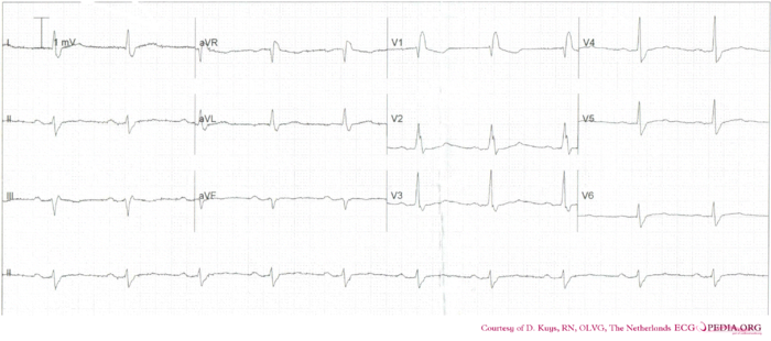
- The ECG starts with a regular rhythm with normal P waves (positive in I, III and AVF, negative in AVR), followed by QRS complexes. Sinusrhythm
- Heart rate
- around 60 bpm
- Conduction (PQ,QRS,QT)
- PQ: 240ms QRS: 120ms QT: 440ms QTc: same as QT at this heart rate
- Heartaxis
- Negative in II, III and AVF: left heart axis
- P wave morphology
- The P wave duration is somewhat prolonged.
- QRS morphology
- Wide QRS complexes with [[[RBBB|right bundle branch block]]] pattern. No LVH or pathologic Q waves.
- ST morphology
- ST depression in V1. Overall flat ST segments.
- Compare with the old ECG (not available, so skip this step)
- Conclusion?
- Rhythm
Trifascicular block with first degree AV block, right bundle branch block and left anterior fascicular block.