Answer MI 22

- Following the 7+2 steps:
- Rhythm
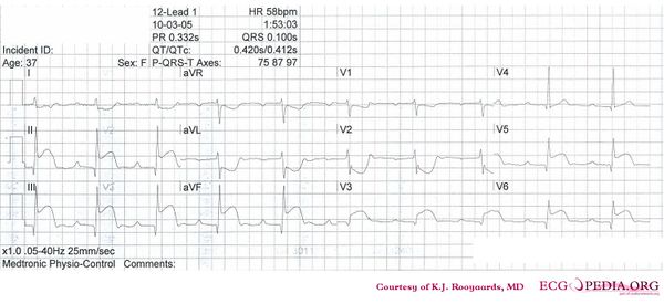
- The ECG shows a regular rhythm with normal P waves (positive in I, III and AVF, negative in AVR), followed by QRS complexes. Sinusrhythm
- Heart rate
- 60 bpm
- Conduction (PQ,QRS,QT)
- PQ: 260ms QRS: 100ms QT: 420ms QTc: 420ms
- Heartaxis
- QRS positive in I and AVF: normal heart axis
- P wave morphology
- The P waves have normal morphology.
- QRS morphology
- Narrow QRS. No left ventricular hypertrophy. Narrow Q waves in lead II,III, AVF, V5 and V6.
- ST morphology
- Pronounced ST elevation in II, III and AVF and V5 and V6. Also ST elevation in V3 which is located at the V4R position. Pronounced ST depression in I, AVL, V1-V2
- Compare with the old ECG (not available, so skip this step)
- Conclusion?
- Rhythm
Sinusbradycardia with first degree AV block and inferior-posterior-lateral myocardial infarction. Lead V4R is clearly elevated, which strongly suggests occlusion of the right coronary artery (RCA).