Answer 2008 05
| Author(s) | A.A.M. Wilde | |
| NHJ edition: | 2008:05,175 | |
| These Rhythm Puzzles have been published in the Netherlands Heart Journal and are reproduced here under the prevailing creative commons license with permission from the publisher, Bohn Stafleu Van Loghum. | ||
| The ECG can be enlarged twice by clicking on the image and it's first enlargement | ||


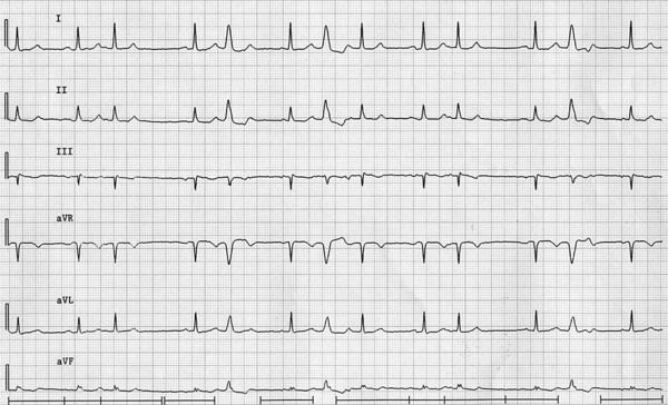
A 55-year-old male without cardiac history is complaining of an irregular heartbeat. Physical examination and an echo reveal no abnormalities. His ECG (extremity leads only) is shown in figure 1.
What is your diagnosis?
Answer
The ECG depicted shows an irregular heart rhythm based on extrasystoles. Some of them have a narrow QRS complex (third and tenth QRS complex) and some have a wide QRS complex (fifth, seventh and twelfth). The basic supraventricular rhythm seems to be sinus rhythm with positive P waves in leads I and II and a negative P wave in lead aVR. The absence of a clear positive P wave in the aVF lead should raise the possibility of an alternative background rhythm (i.e. from a lower placed ectopic atrial focus).
The third QRS complex is followed by a P wave with a different configuration (see lead III, for example). The PQ interval is slightly longer, which is not unusual in the presence of an atrial extrasystole. The fifth QRS complex has an LBBB morphology and is not preceded by a clear P wave. This does not rule out atrial ectopy with aberrant conduction because the P wave might be buried somewhere in the ST segment. It is preceded by a relatively long pause, which based on the PP interval should be referred to as an incomplete compensatory pause. Indeed, aberrant conduction is more prevalent after a longer preceding RR interval (i.e. Ashman phenomenon, see the previous rhythm puzzle. The alternative diagnosis, however, is a ventricular extrasystole. The next QRS complex (seventh QRS complex) is wide again, but this time not preceded nor followed by a pause. In fact the atrial (sinus) rhythm seems undisturbed. This phenomenon is referred to as an interpolated extrasystole and could be considered almost impossible for an atrial extrasystole. Atrial input almost always intervenes with sinus rhythm giving rise to an incomplete compensatory pause (see the pause after the third and tenth QRS complexes). Ventricular extrasystoles can lead to an incomplete compensatory pause (when retrogradely conducted to the atrium, as might be the case for both the fifth and seventh extrasystoles), to a complete compensatory pause (when not retrogradely conducted) and rarely can be interpolated, not disturbing the atrial rhythm.
Although at first glance the wide QRS complexes, in the presence of atrial extrasystoles with narrow QRS complexes, seem to be aberrantly conducted atrial extrasystoles, the most likely diagnosis is ventricular extrasystoles.