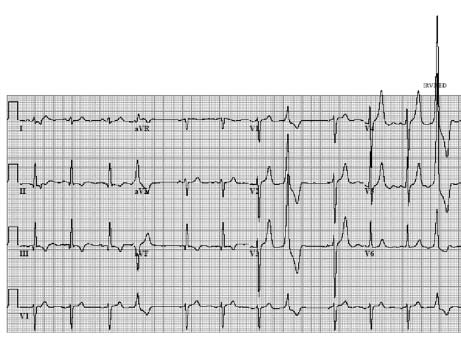
Where do the extras come from
| Author(s) | A.A.M. Wilde, R.B.A. van den Brink | |
| NHJ edition: | 2005:2,67 | |
| These Rhythm Puzzles have been published in the Netherlands Heart Journal and are reproduced here under the prevailing creative commons license with permission from the publisher, Bohn Stafleu Van Loghum. | ||
| The ECG can be enlarged twice by clicking on the image and it's first enlargement | ||

A 64-year-old man had an inferior myocardial infarction ten years ago. Lately he has been having palpitations with occasional dizziness. He sought the attention of his cardiologist. Physical examination revealed no particular abnormalities with the exception of a laterally displaced ictus cordis. His 12-lead ECG, shown in figure 1, was in sinus rhythm with some extrasystoles. The electrical axis is vertical and the Q waves and abnormal ST-T segments in the inferior leads are compatible with an old inferior myocardial infarction. An echocardiogram revealed reduced left ventricular function with extended inferior wall akinesia. The left ventricular ejection fraction was estimated to be 30%.
The questions to address are: what is the meaning of the extrasystoles, where do they come from and should further investigations be performed?