Supraventricular Rhythms
Narrow complex tachycardias
Atrial flutter
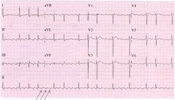
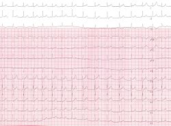
During atrial flutter the atria depolarize in an organized circular movement. This is caused by re-entry. The atria contract typically at around 300 bpm, which results in a fast sequence of p-waves in a sawtooth pattern on the ECG. For most AV-nodes this is way to fast to be able to conduct the signal to the ventricles, so typically there is a 2:1, 3:1 or 4:1 block, resulting in a ventricular frequency of 150, 100 or 75 bpm respectively. Often the grade of block changes every couple of beats, resulting in e.g. 2:1, or 3:1 blocks and a somewhat irregular ventricular heart rate. The saw-tooth is especially prominent in lead II, this lead normally shows constant electrical activity: it is never horizontal. Causes and risk of atrial flutter are comparable to atrial fibrillation.
Atrial fibrillation
Main article: Atrial Fibrillation
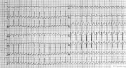
During atrial fibrillation the atria show chaotic depolarisation with multiple foci. Mechanically the atria stop contracting after several days to weeks of atrial fibrillation, the result of the ultra-rapid depolarisations that occur in the atria, typically around 400 bpm, but up to 600 bpm. At the AV node 'every now and then' a beat is conducted to the ventricles, resulting in an irregular ventricular rate, which is the typical ECG characteristic of atrial fibrillation. Sometimes atrial fibrillation results in a course atrial flutter wave on the ECG, but the baseline can also be flat. A flat baseline is more often seen in long standing atrial fibrillation. The cardiac stroke volume is reduced by 10-20% during atrial fibrillation, as the 'atrial kick' is missing and because the heart does not have time to fill at the often higher ventricular rate. Causes; age (+- 10% of 70+ year olds and 15% of 90+ year olds have AFIB kelley), ischemia, hyperthyreoidism, alcohol abuse. Risc: thrombo-embolisation of thrombi that form in the atrial caverns as a result of the reduced atrial motion. These thrombi can emblise to the brain and cause strokes.
Atrial flutter can be catechorized as follows:
- First documented episode:
- Recurrent atrial fibrillation: after two or more episodes.
- Paroxysmal atrial fibrillation: if recurrent atrial fibrillation spontaneously converts to sinus rhythm.
- Persisting atrial fibrillation: if an episode of atrial fibrillation persists more than 7 days.
- Permanent atrial fibrillation: if atrial fibrillation persists after an effort of electrical or chemical cardioversion
Lone AF is atrial fibrillation in patients younger than 60 years in whom no clinical or electrocardiographic signs of heart or lung disease are present. These patiens have a favourable prognosis regarding thrombo-embolic events.
Non-valvular atrial fibrillation is atrial fibrillation in patients without heart valve disease or heart valve replacement or repair. ESCAF
Atrial Tachycardia
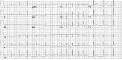
Atrial tachcyardia is a more or less regular heart rate > 100 bpm that does not origin from the sinus node. The p-waves therefore have a different configuration and easily be recognized if the are negative in AVF.
AVNRT
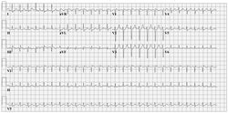
An AV Nodal Re-entry Tachycardia (AVNRT) is a rapid tachycardia with a typical frequency around 200 bpm. The tachycardia origin is the AV node. A prerequisite for AVNRT is a slow and fast pathway in the AV node, most often caused by degradation of the AV nodular tissue. The dual pathways facilitate re-entry.
Two sensitive characteristics to identify AVNRT on the ECG are:
- R'. This is a small secondary R wave. It resembles a right bundel branch block, but the QRS width stays < 120ms.
- RP << 100ms. The distance between the R and P waves is less than 100ms.
Atrio-ventricular Reentry Tachycardia AVRT
Atrio-ventricular Re-entry Tachycardias are somewhat similar to AVNRT. An important diffence however is that an accessory bundle is present in AVRT. This accessory bundle connects the atria and ventricles, thereby bypassing the AV node. The most common type of accessory bundle is a bundle of Kent. AVRTs can result from abnormal retrograde conduction (from ventricles to the atria) and anterorade conduction (from atria to ventricles). This results in two type of circular tachycardias:
- Orthodrome AV Re-entry Tachcardia (conduction retrogarde through the accessory bundle). Usually a narrow QRS complex preceded by a p-wave.
- Antidrome Atrioventricular Re-entry Tachcardia (anterogarde conduction through the abnormal accessory bundle). The ECG shows wide QRS complexes followed by retrograde P-waves. The RP-time is >> 100ms.
- Concealed Bypass Tract
AV junctional tachycardia
