Intraventricular Conduction
| Author(s) | J.S.S.G. de Jong, MD | |
| Moderator | J.S.S.G. de Jong, MD | |
| Supervisor | ||
| some notes about authorship | ||
Conduction delay
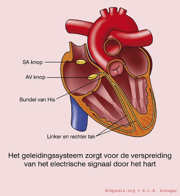
If the QRS complex is wider than 0.12 seconds this is mostly caused by a delay in the conduction tissue of on of the bundle branches:
- Left Bundle Branch Block (LBTB))
- Right Bundle Branch Block(RBTB)
- Interventricular conduction delay
A right or left axis rotation can be caused by a:
Sometimes this conduction delay is frequency-depending: the bundle branch block occurs only at higher heart rates and disappears at slower heart rates.
LBTB vs RBTB
Watch V1 if QRS > 0,12 sec.
If the last QRS activity in V1 is below the baseline (moving away from V1), than LBTB is the most likely diagnosis.
If the last activity is above the baseline, it's a RBTB
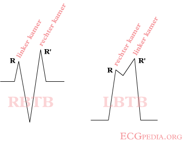
If the QRS > 0.12 sec. but you can't tell whether it's a RBTB or LBTB, than it's 'interventriculair conduction delay', which is allways right. If the conduction system is deteriorates further, the conduction can be taken over by the cardiomyocytes. The conduct the electrical signal cell-to-cell which results in a very wide QRS complex (> 0.16-0.20 seconds).
LBTB
Left bundle branch block
QRS >0,12 sec met brede R in I aVL V5V6 en afwezige q aldaar.
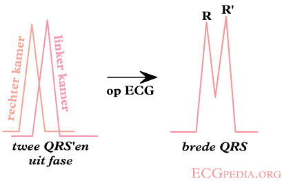
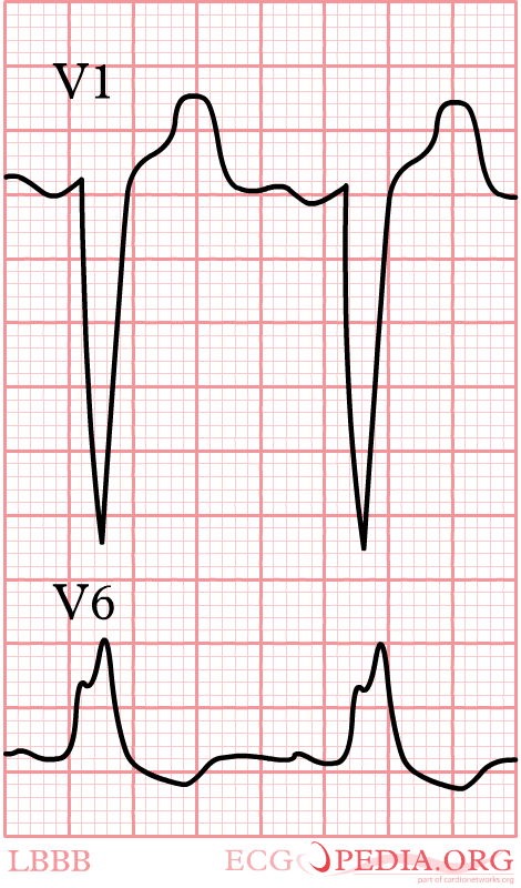
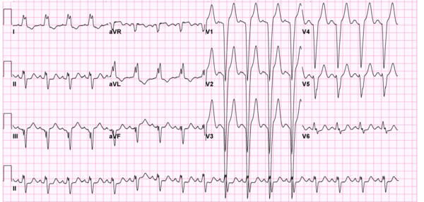
Bij een linker bundeltakblok (LBTB), is de geleiding door de linker bundel vertraagd. Het begin van de depolarisatie is normaal, maar de laterale wand van de linker ventrikel depolariseert dus sterk vertraagd. Hierdoor is er nog electrische activiteit in de linker ventrikel op het moment dat de rest van het hart al 'klaar' is, deze wordt dus niet meer geneutraliseerd door de rechter ventrikel. De laatste activiteit gaat dus naar links, ofwel van V1 af. Met deze kennis is een LBTB makkelijk te begrijpen. Het resultaat ziet er als volgt uit:
Rechter bundeltakblok
QRS >0,12 sec. met RSR'-patroon in V1V2 waarbij R' >R.
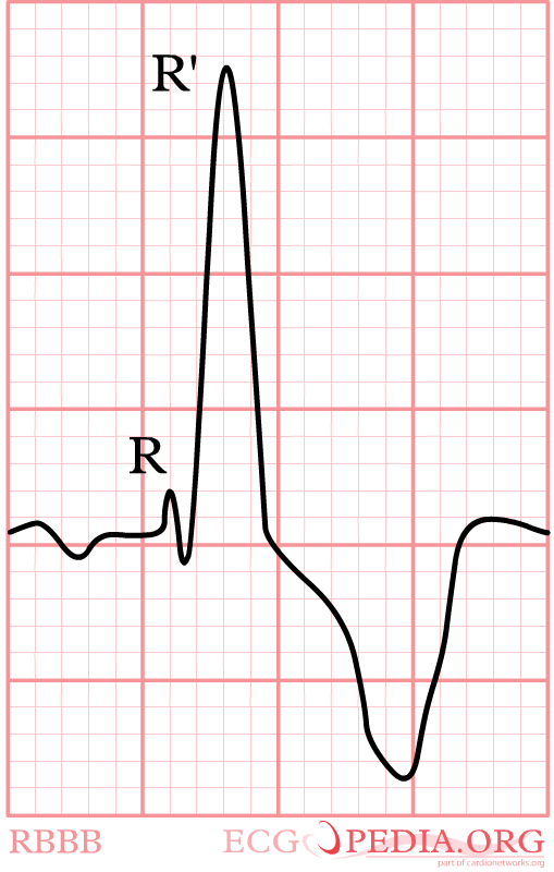

Door in V1 te kijken, kan je deze opties onderscheiden.
Bij een rechter bundeltakblok (RBTB) is het precies andersom. Er is nog electrische activiteit in de rechter ventrikel, terwijl de rest van het hart al 'klaar' is. De laatste activiteit gaat dus naar rechts, ofwel naar V1 toe.
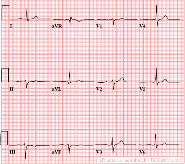
Criteria voor LAFB
Bij een linker anterior fascie blok is de voorste bundel van de twee linker bundels geblokkeerd. Hierdoor wordt de voorwand als laatste ontladen. Dit uit zich in een linker asdraai. De QRS-duur is <0,12 seconde.
asdeviatie naar links (<-30°);
geen of vrijwel geen S in I alwaar normale kleine q;
S >R in II, III;
QRS niet of slechts in geringe mate verbreed.
Criteria voor LPFB
Criteria voor een posterior fascie blok:
asdeviatie naar rechts >+120°;
diepe S in I;
kleine q in III;
QRS niet of slechts in geringe mate verbreed;
criteria voor RVH of oud lateraal myocardinfarct mogen niet aanwezig zijn.