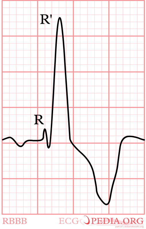
RBBB
- Criteria for right bundle branch block (RBBB)
- QRS >0,12 sec
- Slurred S wave in lead I and V6
- RSR'-pattern in V1 where R' > R


Again, watch V1. In right bundle branch block (RBBB) the conduction in the bundle to the right ventricle is slow. As the right ventricles depolarizes, the left ventricle is often halfway finished and few counteracting electrical activity is left. The last electrical activity is thus to the right, or towards lead V1. In RBBB the QRS complex in V1 is always markedly positive. RBBB is a common finding in healthy individuals. In a recent analysis of 43401 military conscripts, 13.5% had RBBB rbbb
Diagnosing myocardial infarction in RBBB is not as difficult as in LBBB.
More specific definitions
A more specific definition of RBBB is given by the ACC/ESC consensus documentESC-ECG:
Complete RBBB
- QRS duration greater than or equal to 120 ms in adults, greater than 100 ms in children ages 4 to 16 years, and greater than 90 ms in children less than 4 years of age.
- rsr′, rsR′, or rSR′ in leads V1 or V2. The R′ or r′ deflection is usually wider than the initial R wave. In a minority of patients, a wide and often notched R wave pattern may be seen in lead V1 and/or V2.
- S wave of greater duration than R wave or greater than 40 ms in leads I and V6 in adults.
- Normal R peak time in leads V5 and V6 but greater than 50 ms in lead V1.
Of the above criteria, the first 3 should be present to make the diagnosis. When a pure dominant R wave with or without a notch is present in V1, criterion 4 should be satisfied.
Incomplete RBBB
Incomplete RBBB is defined by QRS duration between 110 and 120 ms in adults, between 90 and 100 ms in children between 4 and 16 years of age, and between 86 and 90 ms in children less than 8 years of age. Other criteria are the same as for complete RBBB. In children, incomplete RBBB may be diagnosed when the terminal rightward deflection is less than 40 ms but greater than or equal to 20 ms. The ECG pattern of incomplete RBBB may be present in the absence of heart disease, particularly when the V1 lead is recorded higher than or to the right of normal position and r′ is less than 20 ms.
The terms rsr′ and normal rsr′ are not recommended to describe such patterns, because their meaning can be variously interpreted. In children, an rsr′ pattern in V1 and V2 with a normal QRS duration is a normal variant.
{{{1}}}