Answer MI 19

- Following the 7+2 steps:
- Rhythm
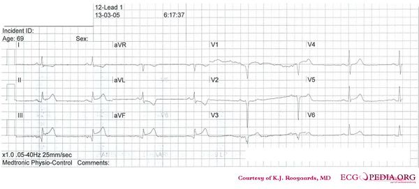
- Regular rhythm with normal P waves (positive in I, II, negative in AVR), followed by QRS complexes. Sinusrhythm
- Heart rate
- 46 bpm. Thus sinusbradycardia
- Conduction (PQ,QRS,QT)
- PQ: 170ms QRS: 110ms QT: 420ms QTc: 370ms
- Heartaxis
- Positive in I and AVL: normal heart axis
- P wave morphology
- P wave positive in II, bifasic in V1. Not well discernible in AVF. Seems normal.
- QRS morphology
- Pathologic Q waves in III and AVF. Slow R wave progression. Some aspecific intraventricular conduction delays (QRS 110ms).
- ST morphology
- ST elevation in II, III (III>II), AVF, V5 (1 mm elevation V4 and V6). ST depression in I and AVL. Lead V3 shows V4R and is not elevated.
- Compare with the old ECG (not available, so skip this step)
- Conclusion?
- Rhythm
Inferior myocardial infarction with Q waves in the inferior leads; probably due to RCA occlusion (III>II, depression in I)