A Concise History of the ECG


The history of the ECG goes back more than one and a half century
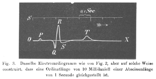
In 1843 Emil Du Bois-Reymond, a german physiologist, was the first to describe "action potentials" of muscular contraction. He used a highly sensitive galvanometer, which contained more than 5 km of wire. Du Bios Reymond named the different waves: "o" was the stable equilibrium and he was the first to use the p, q, r and s to describe the different waves. Dubois However, in his excellent paper on the 'Naming of the waves in the ECG' Dr Hurst credits Einthoven for being the first to use PQRS and T.Hurst
In 1850 M. Hoffa described how he could induce irregular contractions of the ventricles of doghearts by administering electrical shock. Hoffa
In 1887 the English physiologist Augustus D. Waller from Londen published the first human electrocardiogram. He used a capillar-electrometer. Waller
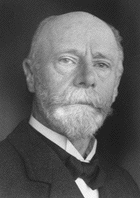
The dutchman Willem Einthoven (1860-1927) introduced in 1893 the term 'electrocardiogram'. He described in 1895 how he used a galvanometer to visualize the electrical activity of the heart. In 1924 he received the Nobelprize for his work on the ECG. He connected electrodes to a patienta showed the electrical difference between two electrodes on the galvanometer. We still now use the term: Einthovens'leads. The string galvanometer (see Image) was the first clinical instrument on the recording of an ECG.
In 1905 Einthoven recorded the first 'telecardiogram' from the hospital to his laboratoy 1.5 km away.
In 1906 Einthoven published the first article in which he described a series of abnormal ECGs: left- and right bundlebranchblock, left- and right atrialdilatation, the U wave, notching of the QRS complex, ventricular extrasystoles, bigemini, atrialflutter and total AV block. Einthoven