ECG in Congenital Heart Disease
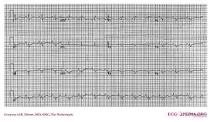
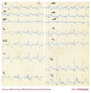
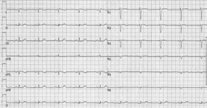
Congenital Heart Disease can result in ECG changes, often related to atrial or ventricular overload and enlargement. Below a list of relatively common forms of congenital heart disease and their potential ECG changes. Adapted from Khairy et al.khairy
Secundum atrial septal defect
Information about Secundum atrial septal defec on Wikipedia (external link)
- Rhythm: normal sinus rhythm, increased risk of AF with age
- PR interval: first degree AV block in 6-19%
- QRS axis: 0° to 180°; RAD; LAD in Holt-Oram or LAHB
- QRS Configuration: rSr´ or rsR´ with RBBBi>RBBBc
- Atrial Enlargement: RAE 35%
- Ventricular hypertrophy: Uncommon
- Particularities: "Crochetage" pattern
Ventricular Septal Defect
- Rhythm: normal sinus rhythm, PVCs
- PR interval: Normal or mild ↑; 1° AVB 10%
- QRS axis: RAD with BVH; LAD 3% to 15%
- QRS Configuration: Normal or rsr´; possible RBBB
- Atrial Enlargement: Possible RAE±LAE
- Ventricular hypertrophy: BVH 23% to 61%; RVH with Eisenmenger
- Particularities: Katz-Wachtel phenomenon
AV canal defect
- Rhythm: normal sinus rhythm, PVCs 30%
- PR interval: 1° AVB >50%
- QRS axis: Moderate to extreme LAD; normal with atypical
- QRS Configuration: rSr´ or rsR´
- Atrial Enlargement: Possible LAE
- Ventricular hypertrophy: Uncommon in partial; BVH in complete; RVH with Eisenmenger
- Particularities: Inferoposteriorly displaced AVN
Patent ductus arteriosus
- Rhythm: normal sinus rhythm, ↑ IART/AF with age
- PR interval: ↑ PR 10% to 20%
- QRS axis: Normal
- QRS Configuration: Deep S V1, tall R V5 and V6
- Atrial Enlargement: LAE with moderate PDA
- Ventricular hypertrophy: Uncommon
- Particularities: Often either clinically silent or Eisenmenger
Pulmonary stenosis
- Rhythm: normal sinus rhythm
- PR interval: Normal
- QRS axis: Normal if mild; RAD with moderate/severe
- QRS Configuration: Normal; or rSr´; R´ increases with severity
- Atrial Enlargement: Possible RAE
- Ventricular hypertrophy: RVH; severity correlates with R:S in V1 and V6
- Particularities: Axis deviation correlates with RVP
Aortic coarctation
- Rhythm: normal sinus rhythm
- PR interval: Normal
- QRS axis: Normal or LAD
- QRS Configuration: Normal
- Atrial Enlargement: Possible LAE
- Ventricular hypertrophy: LVH, especially by voltage criteria
- Particularities: Persistent RVH rare beyond infancy
Ebstein’s anomaly


Information about Ebstein's anomaly on Wikipedia (external link)
- Rhythm: normal sinus rhythm, possible EAR, SVT; AF/IART 40%
- PR interval: 1° AVB common; short if WPW
- QRS axis: Normal or LAD
- QRS Configuration: Low-amplitude multiphasic atypical RBBB
- Atrial Enlargement: RAE with Himalayan P waves
- Ventricular hypertrophy: Diminutive RV
- Particularities: Accessory pathway common; Q II, III, aVF, and V1–V4
Surgically repaired TOF
- Rhythm: normal sinus rhythm, PVCs; IART 10%; VT 12%
- PR interval: Normal or mild ↑
- QRS axis: Normal or RAD; LAD 5% to 10%
- QRS Configuration: RBBB 90%
- Atrial Enlargement: Peaked P waves; RAE possible
- Ventricular hypertrophy: RVH possible if RVOT obstruction or PHT
- Particularities: QRS duration±QTd predictive of VT/SCD
Congenitally corrected TGA
- Rhythm: normal sinus rhythm
- PR interval: 1° AVB >50%; AVB 2%/year
- QRS axis: LAD
- QRS Configuration: Absence septal q; Q in III, aVF, and right precordium
- Atrial Enlargement: Not if no associated defects
- Ventricular hypertrophy: Not if no associated defects
- Particularities: Anterior AVN; positive T precordial; WPW with Ebstein’s
Complete TGA/intra-atrial baffle
- Rhythm: Sinus brady 60%; EAR; junctional; IART 25%
- PR interval: Normal
- QRS axis: RAD
- QRS Configuration: Absence of q, small r, deep S in left precordium
- Atrial Enlargement: Possible RAE
- Ventricular hypertrophy: RVH; diminutive LV
- Particularities: Possible AVB if VSD or TV surgery
UVH with Fontan
- Rhythm: Sinus brady 15%; EAR; junctional; IART >50%
- PR interval: Normal in TA; 1° AVB in DILV
- QRS axis: LAD in single RV, TA, single LV with noninverted outlet
- QRS Configuration: Variable; ↑R and S amplitudes in limb and precordial leads
- Atrial Enlargement: RAE in TA
- Ventricular hypertrophy: RVH with single RV; possible LVH with single LV
- Particularities: Absent sinus node in LAI; AV block with L-loop or AVCD
Dextrocardia

- Rhythm: normal sinus rhythm, P-wave axis 105° to 165° with situs inversus
- PR interval: Normal
- QRS axis: RAD
- QRS Configuration: Inverse depolarization and repolarization
- Atrial Enlargement: Not with situs inversus
- Ventricular hypertrophy: LVH: tall R V1–V2; RVH: deep Q, small R V1 and tall R right lateral
- Particularities: Situs solitus: normal P-wave axis and severe CHD
ALCAPA
- Rhythm: normal sinus rhythm
- PR interval: Normal
- QRS axis: Possible LAD
- QRS Configuration: Ant-lat Q waves; possible ant-sept Q waves
- Atrial Enlargement: Possible LAE
- Ventricular hypertrophy: Selective hypertrophy of posterobasal LV
- Particularities: Possible ischemia
References
<biblio>
- khairy pmid=18056539
</biblio>