Syncopated Rhythm: Difference between revisions
Jump to navigation
Jump to search
mNo edit summary |
m moved Syncopated rhythm to Syncopated Rhythm |
(No difference)
| |
Latest revision as of 20:09, 25 January 2010
| Author(s) | L.R.C. Dekker, R. Tukkie | |
| NHJ edition: | 2007:4,157 | |
| These Rhythm Puzzles have been published in the Netherlands Heart Journal and are reproduced here under the prevailing creative commons license with permission from the publisher, Bohn Stafleu Van Loghum. | ||
| The ECG can be enlarged twice by clicking on the image and it's first enlargement | ||

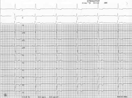
A 65-year-old woman was admitted because of recurrent syncope. Her complaints were difficult to interpret due to mental impairment after cerebral haemorrhage ten years earlier. She was once found lying in the street after walking her dog. Her medical history further included congestive heart failure after several untreated myocardial infarctions and diabetes. Under treatment with oral antidiabetics, furosemide, ACE inhibitor, digoxin and aspirin, she was in functional NYHA class I/IV. A 12-lead ECG is shown in figure 1.
What is the most likely level of this 2:1 AV block and what explains the p-wave pattern?