Ventricular Flutter: Difference between revisions
Jump to navigation
Jump to search
mNo edit summary |
No edit summary |
||
| Line 12: | Line 12: | ||
| example2 = | | example2 = | ||
}} | }} | ||
[[Image:ventricular_flutter_12lead.jpg|thumb|A ventricular flutter on a 12 lead ECG]] | |||
Ventricular Flutter is mostly caused by re-entry with a frequency of 300 bpm. The ECG shows a typical sinusoidal pattern. During ventricular flutter the ventricles depolarize in a circular pattern, which prevents good function. Most often this results in a minimal cardiac output and subsequent ischemia. Often deteriorates into [[Ventricular Fibrillation]].{{clr}} | Ventricular Flutter is mostly caused by re-entry with a frequency of 300 bpm. The ECG shows a typical sinusoidal pattern. During ventricular flutter the ventricles depolarize in a circular pattern, which prevents good function. Most often this results in a minimal cardiac output and subsequent ischemia. Often deteriorates into [[Ventricular Fibrillation]].{{clr}} | ||
Latest revision as of 15:41, 2 February 2008
| This is part of: Ventricular Arrhythmias |

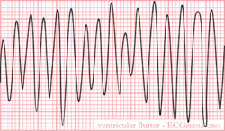
Ventricular Flutter is mostly caused by re-entry with a frequency of 300 bpm. The ECG shows a typical sinusoidal pattern. During ventricular flutter the ventricles depolarize in a circular pattern, which prevents good function. Most often this results in a minimal cardiac output and subsequent ischemia. Often deteriorates into Ventricular Fibrillation.
