File:Brugada syndrome type1 example1.png: Difference between revisions
Jump to navigation
Jump to search
No edit summary |
|||
| Line 1: | Line 1: | ||
== Summary == | == Summary == | ||
{{Information | {{Information | ||
|Description = Brugada syndrome type 1. Note that V5 is placed one intercostal space above V1 (V1 IC4) and V6 is placed one intercostal space above V2 (V2 IC3). Type I morphology is seen in V1, V1 IC3 and V2 IC3. Furthermore, there is a horizontal QRS axis, broad P waves, wide S waves in the lateral and inferior leads and fractionation of the QRS complex in III and aVL. | |Description = '''Brugada syndrome type 1'''. Note that V5 is placed one intercostal space above V1 (V1 IC4) and V6 is placed one intercostal space above V2 (V2 IC3). Type I morphology is seen in V1, V1 IC3 and V2 IC3. Furthermore, there is a horizontal QRS axis, broad P waves, wide S waves in the lateral and inferior leads and fractionation of the QRS complex in III and aVL. | ||
|Source = P.G. Postema, MD | |Source = P.G. Postema, MD | ||
|Date = 2007 | |Date = 2007 | ||
Latest revision as of 21:57, 2 December 2007
Summary
| Description |
Brugada syndrome type 1. Note that V5 is placed one intercostal space above V1 (V1 IC4) and V6 is placed one intercostal space above V2 (V2 IC3). Type I morphology is seen in V1, V1 IC3 and V2 IC3. Furthermore, there is a horizontal QRS axis, broad P waves, wide S waves in the lateral and inferior leads and fractionation of the QRS complex in III and aVL. |
|---|---|
| Category |
{{{Category}}} |
| Source |
P.G. Postema, MD |
| Date |
2007 |
| Author | |
| Permission |
Creative Commons Attribution Noncommercial Share-Alike License |
File history
Click on a date/time to view the file as it appeared at that time.
| Date/Time | Thumbnail | Dimensions | User | Comment | |
|---|---|---|---|---|---|
| current | 10:08, 10 April 2010 | 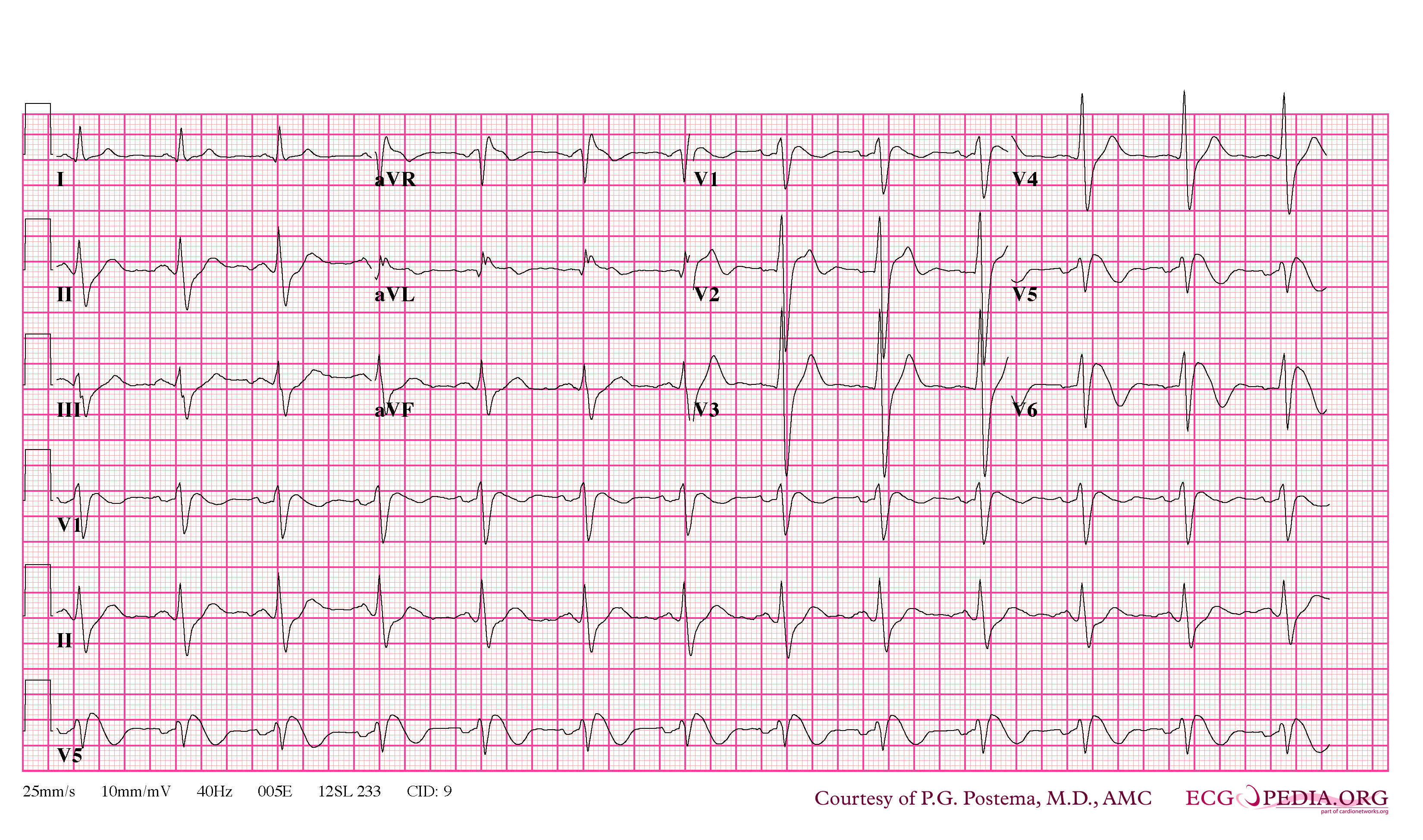 | 3,300 × 1,949 (370 KB) | (username removed) |
File usage
The following page uses this file: