Case 100: Difference between revisions
Jump to navigation
Jump to search
mNo edit summary |
mNo edit summary |
||
| Line 1: | Line 1: | ||
Look at the consecutive ECGs in this patient. What is the problem? | Look at the consecutive ECGs in this patient. What is the problem that causes the arrhythmia in rhythm strip 2? | ||
[[Image:KJcasu17-1.jpg|thumb | [[Image:KJcasu17-1.jpg|thumb| Rhythm strip 1]] | ||
{{clr}} | {{clr}} | ||
[[Image:KJcasu17-2.jpg|thumb | [[Image:KJcasu17-2.jpg|thumb| Rhythm strip 2]] | ||
{{clr}} | {{clr}} | ||
[[Image:KJcasu17-3.jpg|thumb | [[Image:KJcasu17-3.jpg|thumb| Rhythm strip 3]] | ||
{{clr}} | {{clr}} | ||
[[Image:KJcasu17-4.jpg|thumb | [[Image:KJcasu17-4.jpg|thumb| Rhythm strip 4]] | ||
{{clr}} | {{clr}} | ||
| Line 12: | Line 12: | ||
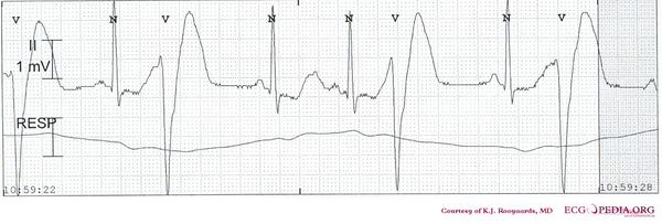
* The first rhythm strip shows sinusrhythm with a severely prolonged QT interval. In total four ventricular premature beats are present on this strip. All fall within the T wave of the previous sinus beat. | * The first rhythm strip shows sinusrhythm with a severely prolonged QT interval. In total four ventricular premature beats are present on this strip. All fall within the T wave of the previous sinus beat. | ||
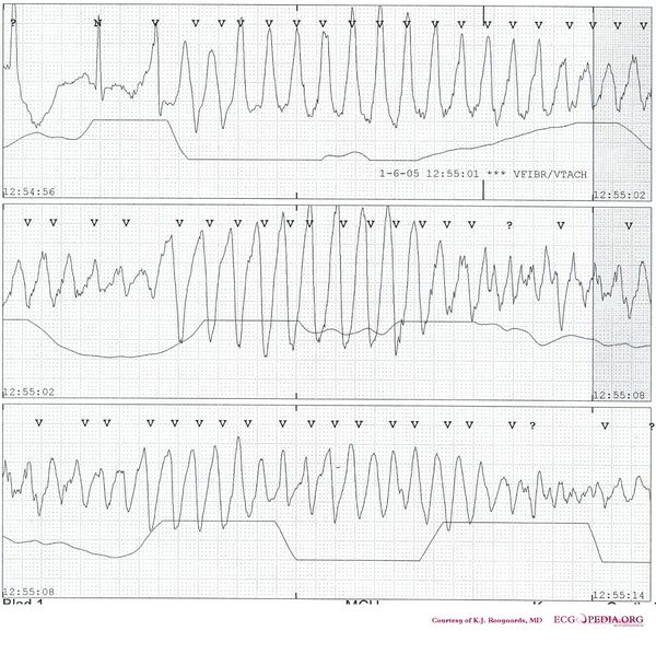
* The second strip shows [[Torsade de Pointes]] with a typical short (not shown) - long - short initial sequence. | * The second strip shows [[Torsade de Pointes]] with a typical short (not shown) - long - short initial sequence. | ||
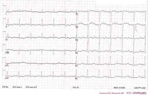
* The | * The third ECG shows severe QT prolongation with a stretched QT segment and small T wave, consistent with Type 2 [[LQTS]]. | ||
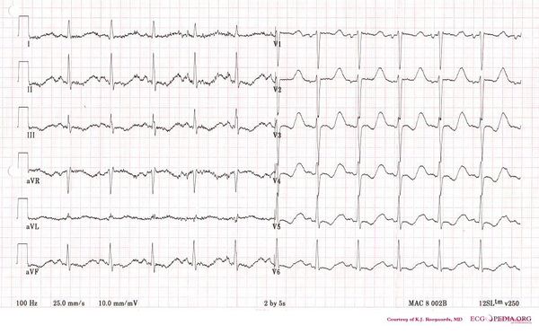
* The fourth ECG shows the same patient later in time. | |||
{{clr}} | {{clr}} | ||
Revision as of 09:59, 29 November 2007
Look at the consecutive ECGs in this patient. What is the problem that causes the arrhythmia in rhythm strip 2?


Answer
- The first rhythm strip shows sinusrhythm with a severely prolonged QT interval. In total four ventricular premature beats are present on this strip. All fall within the T wave of the previous sinus beat.
- The second strip shows Torsade de Pointes with a typical short (not shown) - long - short initial sequence.
- The third ECG shows severe QT prolongation with a stretched QT segment and small T wave, consistent with Type 2 LQTS.
- The fourth ECG shows the same patient later in time.