Inferior MI: Difference between revisions
Jump to navigation
Jump to search
mNo edit summary |
mNo edit summary |
||
| Line 1: | Line 1: | ||
{{Chapter|Myocardial Infarction}} | {{Chapter|Myocardial Infarction}} | ||
'''ST elevation in II, III and aVF''' | '''ST elevation in II, III and aVF''' | ||
[[image:V4R_occlusion.svg|thumb|ST elevation or depression in V4R can help in differentiating a RCA from a RCX occlusion.]] | |||
This part of the heart muscle lies on the diaphragm and is supplied of blood bij the right coronary artery (RCA) in 80% of patients. In the remaing 20% the inferior wall is supplied by the ramus circumflexus(RCX). | This part of the heart muscle lies on the diaphragm and is supplied of blood bij the right coronary artery (RCA) in 80% of patients. In the remaing 20% the inferior wall is supplied by the ramus circumflexus(RCX). | ||
Latest revision as of 09:51, 14 October 2007
| This is part of: Myocardial Infarction |
ST elevation in II, III and aVF

This part of the heart muscle lies on the diaphragm and is supplied of blood bij the right coronary artery (RCA) in 80% of patients. In the remaing 20% the inferior wall is supplied by the ramus circumflexus(RCX).
An occlusion of the RCA can be distinguished of a RCX occulusion on the ECG:Zimetbaum
- Distal RCA occlusion (sens 90%, spec 71%)
- ST segment elevation in III higher than ST segment elevation in II ("the highest elevation points at the culprit")and
- ST segment depression in I, AVL, or both (>1 mm)
- Proximal RCA occlusion (sens 79%, spec 100%)
- Additional ST segment elevation in V1, V4R or both
- RCX occlusion (sens 83%, spec 96%)
- ST segment elevation in I, AVL, V5, and V6 and
- ST segment depression in V1, V2, and V3
Examples
-
![A typical example of an inferior wall infarction.]]](https://nl.ecgpedia.org/images/thumb/a/ac/AMI_inferior.jpg/120px-AMI_inferior.jpg) A typical example of an inferior wall infarction.]]
A typical example of an inferior wall infarction.]] -
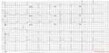 Inferior-posterior MI due to RCA occlusion
 -
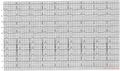 Inferior MI due to RCA occlusion
 -
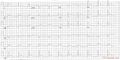 Inferior MI due to RCX occlusion
 -
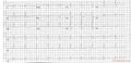 Posterior-lateral MI due to RCX occlusion
![A typical example of an inferior wall infarction.]]](/wiki/File:AMI_inferior.jpg)


References
<biblio>
- Zimetbaum pmid=12621138
</biblio>