McGill Case 136: Difference between revisions
Jump to navigation
Jump to search
No edit summary |
No edit summary |
||
| Line 6: | Line 6: | ||
}} | }} | ||
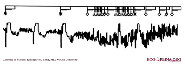
[[File: | [[File:E00031361.jpg|thumb|600px|left|This is an illustration of pectoral muscle pacemaker inhibition. It is particularly interesting as it shows a maker channel to document what the pacemaker is seeing. This patient was totally dependent on his pacer and was loosing consciousness in spite of his pacemaker. | ||
These are two separate strips from the same session. The stars represent paced beats, the diamonds sensed beats, and the diamonds with a cross indicate noise. | |||
Note the EKG records the muscle artifact (noisy baseline) and note the sensing of the pectoral muscle and the loss of paced beats as documented in the marker channel. | |||
The patient's symptoms stopped with reprogramming the pacemaker.]] | |||
[[File:E00031362.jpg|thumb|600px|left|]] | |||
Latest revision as of 19:56, 17 February 2012

|

