P Wave Morphology: Difference between revisions
Jump to navigation
Jump to search
mNo edit summary |
mNo edit summary |
||
| Line 12: | Line 12: | ||
|editor= | |editor= | ||
}} | }} | ||
{{box| | |||
The '''P wave morphology''' can reveal right or left atrial stretch or atrial arrhythmias and is best determined in leads II and V1 during sinus rhythm. | The '''P wave morphology''' can reveal right or left atrial stretch or atrial arrhythmias and is best determined in leads II and V1 during sinus rhythm. | ||
| Line 22: | Line 23: | ||
*The p wave duration is usually shorter than 0.12 seconds | *The p wave duration is usually shorter than 0.12 seconds | ||
|} | |} | ||
}} | |||
==The abnormal P wave== | |||
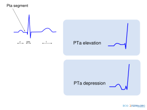
Elevation or depression of the [[PTa segment]] (the part between the p wave and the beginning of the QRS complex) can result from [[Ischemia#Atrial infarction|Atrial infarction]] or [[Clinical Disorders#Pericarditis|pericarditis]]. | Elevation or depression of the [[PTa segment]] (the part between the p wave and the beginning of the QRS complex) can result from [[Ischemia#Atrial infarction|Atrial infarction]] or [[Clinical Disorders#Pericarditis|pericarditis]]. | ||
Revision as of 18:57, 30 May 2009
| «Step 4:Heart axis | Step 6: QRS morphology» |
| Author(s) | J.S.S.G. de Jong, MD, A. Bouhiouf, Msc | |
| Moderator | J.S.S.G. de Jong, MD | |
| Supervisor | ||
| some notes about authorship | ||
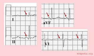
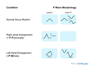
The P wave morphology can reveal right or left atrial stretch or atrial arrhythmias and is best determined in leads II and V1 during sinus rhythm.
{
The abnormal P wave
Elevation or depression of the PTa segment (the part between the p wave and the beginning of the QRS complex) can result from Atrial infarction or pericarditis.
If the p-wave is enlarged, the atria are enlarged.
If the P wave is inverted, it is most likely an ectopic atrial rhythm not originating from the sinus node.
Examples
{{{1}}}


