LBBB: Difference between revisions
Jump to navigation
Jump to search
changed to "can be difficult" -- i will check out "MI Diagnosis in LBBB" before I go on about Sgarbossa on this page |
No edit summary |
||
| Line 6: | Line 6: | ||
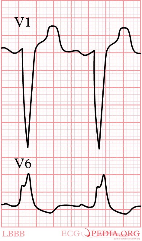
[[Image:LBBB.png|thumb| In a LBBB, the last depolarization wave is in the left ventricle. This wave is directed away from V1. On the ECG, V1 will show a negative complex.]] | [[Image:LBBB.png|thumb| In a LBBB, the last depolarization wave is in the left ventricle. This wave is directed away from V1. On the ECG, V1 will show a negative complex.]] | ||
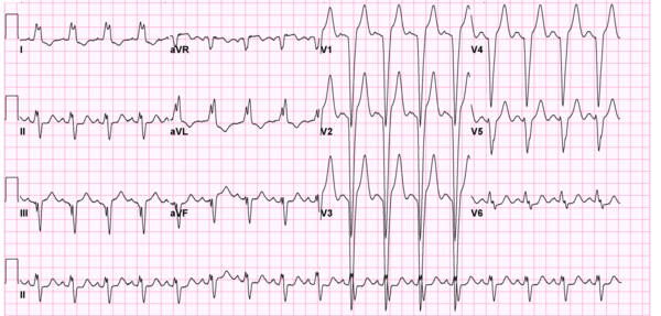
[[Image:12leadLBTB.png|thumb| Left bundle branch Block on a 12 lead ECG.]] | [[Image:12leadLBTB.png|thumb| Left bundle branch Block on a 12 lead ECG.]] | ||
[[Image:12leadLBTB002.jpg|thumb| Another example of Left bundle branch Block on a 12 lead ECG.]] | |||
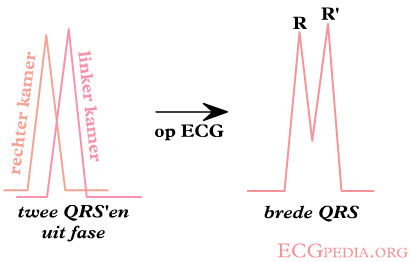
In ''left bundle branch block'' (LBBB) the conduction in the left bundle is slow. This results in delayed depolarization of the left ventricle, especially the left lateral wall. The electrical activity in the left lateral wall is unopposed by the usual right ventricular electrical activity. The last activity on the ECG thus goes to the left or away from V1. Once you remember this, LBBB is easy to understand. | In ''left bundle branch block'' (LBBB) the conduction in the left bundle is slow. This results in delayed depolarization of the left ventricle, especially the left lateral wall. The electrical activity in the left lateral wall is unopposed by the usual right ventricular electrical activity. The last activity on the ECG thus goes to the left or away from V1. Once you remember this, LBBB is easy to understand. | ||
Revision as of 13:27, 30 September 2007
- Criteria for left bundle branch block (LBBB) Garcia
- QRS >0,12 sec
- Broad monomorphic R waves in I and V6 with no Q waves
- Broad monomorphic S waves in V1, may have a small r wave


In left bundle branch block (LBBB) the conduction in the left bundle is slow. This results in delayed depolarization of the left ventricle, especially the left lateral wall. The electrical activity in the left lateral wall is unopposed by the usual right ventricular electrical activity. The last activity on the ECG thus goes to the left or away from V1. Once you remember this, LBBB is easy to understand.
Diagnosis of myocardial infarction in LBBB can be difficult.
Also read right bundle branch block.
References
<biblio>
- Garcia isbn=0763722464
- wellens isbn=9781416002598
</biblio>