Chamber Hypertrophy and Enlargment: Difference between revisions
| Line 22: | Line 22: | ||
==Criteria for right ventricular hypertrophy== | ==Criteria for right ventricular hypertrophy== | ||
[[Image: | [[Image:Rechter_ventrikel_hypertrofie.GIF.GIF|thumb]] | ||
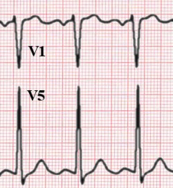
[[Image:E_rvh.jpg|thumb|Right ventricular hypertrohpy, the R wave is greater than the S wave in V1]] | [[Image:E_rvh.jpg|thumb|Right ventricular hypertrohpy, the R wave is greater than the S wave in V1]] | ||
Right ventricular hypertrophy occurs mainly in lung disease or in congenital heart disease. | Right ventricular hypertrophy occurs mainly in lung disease or in congenital heart disease. | ||
Revision as of 15:59, 20 May 2007
In hypertrophy the heart muscle is thicker. This can have different causes. Left ventricular hypertrophy results from an increase in left ventricular workload, e.g. during hypertension or aortic valve stenosis. Right ventricular hypertrophy results from an increase in right ventricular workoad, e.g. emphysema or pulmonary embolisation. These causes are fundamentally different from hypertrophic obstructive cardiomyopathy (HCM), which is a congenital misallignment of cardiomyocytes resulting in hypertrophy.
Left and right ventricular hypertrophy can be distinguished on the ECG:
Criteria for left ventricular hypertrophy


As the left ventricular becomes thicker, the QRS complexes become larger. This is especially true for leads V1-V6. The S wave in V1 is deep, the R wave in V4 is high. Often some ST depression can be seen in leads V5-V6, which is in this setting is called a 'strain pattern'.
To diagnose left ventricular hypertrhophy on the ECG one of the following criteria should be met:
- R in V5 or V6 + S in V1 >35 mm. (this is called the Sokolow-Lyon criterium)
- R >26 mm in V5 or V6;
- R >20 mm in I, II or III;
- R >12 mm in aVL (in the absence of left anterior fascicular block);
The Cornell-criterium has different values in men and women:
- R in aVL and S in V3 >28 mm in men
- R in aVL and S in V3 >20 mm in women
Criteria for right ventricular hypertrophy

Right ventricular hypertrophy occurs mainly in lung disease or in congenital heart disease. The ECG shows a negative QRS complex in I (and thus a right heart axis) and a positive QRS complex in V1.
- R > S in V1 (R must be > 0.5 mV)
- Right heart axis