|
|
| (4 intermediate revisions by the same user not shown) |
| Line 1: |
Line 1: |
| __NOTOC__
| | Below you can find some common examples. ECGs can be magnified by clicking on the image.... |
| <div style="border:1px solid #AAAAAA;padding:4px;text-align:left;"> | | |
| <div style="padding:15px"><div style="font-size:18pt">'''Welcome to ECGpedia'''</div>''a [[w:wiki|wiki]] electrocardiography (ECG) course and textbook designed for medical professionals such as cardiac care nurses and physicians.''</div>
| | __TOC__ |
| <!-- 3 boxes -->
| | {{clr}} |
| <div style="padding:10px"> | | |
| <!-- course boxes -->
| | ==Ischemia & Myocardial Infarction== |
| {| width="100%" cellspacing="5" | | <div align="left"> |
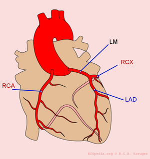
| |- | | [[Image:coronary_anatomy.png|thumb|300px| An overview of the coronary arteries. LM = 'Left Main' = mainstem; LAD = 'Left Anterior Descending' artery; RCX = Ramus Circumflexus; RCA = 'Right Coronary Artery'.]] |
| | style="border:1px solid #E2ACB1;background-color:#FFF5F5;padding:5px;" width="33%" valign="top" | | | |
| <h2 style="margin:0px;margin-bottom:15px;background-color:#D1DAEB;font-size:120%;font-weight:bold;border:1px solid #a3bfb1;text-align:center;color:#000;padding:0.2em 0.4em;">The ECG Course</h2>
| | <div style="width:689px"> |
| [[Image:Course.jpg|140px|center]] | | Guess the culprit coronary artery that was occluded in these examples of myocardial infarctions |
| <div align="center" style="margin-top:15px;">
| | |
| <div style="font-size:10pt;font-weight:bold;border-bottom:1px solid #E2ACB1;color:#000;padding:0.2em 0.4em">Go to the [[ECG course]] for the [[Basics]] and the 7+2 step plan:</div>
| | {{ImageC |image=ami0001.jpg |link=MI 1|text=[[MI 1]]}} |
| {| style="background:transparent;font-size:8pt" align="left" | | {{ImageC |image=ami0002.jpg |link=MI 2|text=[[MI 2]]}} |
| |- | | {{ImageC |image=ami0003.jpg |link=MI 3|text=[[MI 3]]}} |
| | style="background:transparent" valign="top" align="left"| | | {{ImageC |image=ami0004.jpg |link=MI 4|text=[[MI 4]]}} |
| #[[Rhythm]]
| | {{ImageC |image=ami0005.jpg |link=MI 5|text=[[MI 5]]}} |
| #[[Rate]]
| | {{ImageC |image=ami0006.jpg |link=MI 6|text=[[MI 6]]}} |
| #[[Conduction|Conduction (PQ,QRS,QT)]]
| | {{ImageC |image=ami0007.jpg |link=MI 7|text=[[MI 7]]}} |
| #[[Heart axis]]
| | {{ImageC |image=ami0008.jpg |link=MI 8|text=[[MI 8]]}} |
| #[[P wave morphology]]
| | {{ImageC |image=ami0009.jpg |link=MI 9|text=[[MI 9]]}} |
| #[[QRS morphology]]
| | {{ImageC |image=ami0010.jpg |link=MI 10|text=[[MI 10]]}} |
| #[[ST morphology]]
| | {{ImageC |image=ami0011.jpg |link=MI 11|text=[[MI 11]]}} |
| | valign="top" align="left"| | | {{ImageC |image=ami0012.jpg |link=MI 12|text=[[MI 12]]}} |
| #[[Compare_the_old_and_new_ECG|Compare with previous ECG]]
| | {{ImageC |image=ami0013.jpg |link=MI 13|text=[[MI 13]]}} |
| #[[Conclusion]]
| | {{ImageC |image=Casus2_2.jpg |link=MI 14|text=[[MI 14]]}} |
| |} | | {{ImageC |image=AMI_anterior_LAD.jpg |link=MI 15|text=[[MI 15]]}} |
| <div style="clear:both"></div>
| | {{ImageC |image=KJcasus5.jpg |link=MI 16|text=[[MI 16]]}} |
| <div style="font-size:10pt;font-weight:bold;border-top:1px solid #E2ACB1;border-bottom:1px solid #E2ACB1;color:#000;padding:0.2em 0.4em;margin-top:5px;margin-bottom:5px;">Download and Print</div>
| | {{ImageC |image=KJcasus6.jpg |link=MI 17|text=[[MI 17]]}} |
| <div style="border:1px solid #ccc;padding:3px;margin-top:8px;background:white">[[Image:ECG_reference_card_thumbnail.jpg|290px|Click on [http://www.ecgpedia.org/A4/ECGpedia_on_1_A4En.pdf link], not on this image]]</div><div style="font-size:8pt">Download and print our '''[http://www.ecgpedia.org/A4/ECGpedia_on_1_A4En.pdf ECG Reference Card] as PDF''' (new improved version April 2009!, read the [[printing instructions]])</div>
| | {{ImageC |image=KJcasus7.jpg |link=MI 18|text=[[MI 18]]}} |
| <div style="text-align:center;font-size:8pt;border-top:1px solid #E2ACB1;color:#000;padding:0.2em 0.4em;margin-top:5px;">
| | {{ImageC |image=KJcasus8.jpg |link=MI 19|text=[[MI 19]]}} |
| [http://nl.ecgpedia.org/wiki/Powerpoint_presentaties_van_ECG_cursussen Ready-made presentation files for ECG courses (in Dutch)] | | {{ImageC |image=KJcasus10.jpg |link=MI 20|text=[[MI 20]]}} |
| | {{ImageC |image=KJcasus12.jpg |link=MI 21|text=[[MI 21]]}} |
| | {{ImageC |image=KJcasus13.jpg |link=MI 22|text=[[MI 22]]}} |
| | {{ImageC |image=KJcasus16.jpg |link=MI 23|text=[[MI 23]]}} |
| | {{clr}} |
| </div> | | </div> |
| </div> | | </div> |
| <!--start 2nd center box-->
| | {{clr}} |
| |style="border:1px solid #E2ACB1;background-color:#FFF5F5;padding:5px;" width="33%" valign="top" |
| | ==Arrhythmias== |
| <h2 style="margin:0px;margin-bottom:15px;background-color:#D1DAEB;font-size:120%;font-weight:bold;border:1px solid #a3bfb1;text-align:center;color:#000;padding:0.2em 0.4em;">The ECG Textbook</h2>
| | {{ImageC |image=Casus2_2.jpg |link=Case 1|text=[[Case 1]]}} |
| [[Image:book.jpg|140px|center]]
| | {{ImageC |image=KJcasus3.jpg |link=Case 2|text=[[Case 2]]}} |
| <div align="center" style="margin-top:15px;">
| | {{ImageC |image=KJcasus9.jpg |link=Case 3|text=[[Case 3]]}} |
| <div style="font-size:10pt;font-weight:bold;border-bottom:1px solid #E2ACB1;color:#000;padding:0.2em 0.4em">Browse the [[Textbook|ECG Textbook]]:</div>
| | {{ImageC |image=Triblock.png |link=Case 4|text=[[Case 4]]}} |
| {| style="background:transparent;font-size:8pt" align="left" | | {{ImageC |image=JJ00004.jpg |link=Case 5|text=[[Case 5]]}} |
| |- | | {{clr}} |
| | style="background:transparent" valign="top" align="left"|
| | ==Electrolyte Disorders== |
| * [[Normal tracing|Normal tracing]]
| | {{ImageC |image=KJcasu17-1.jpg |link=Case 100|text=[[Case 100]]}} |
| * [[A Concise History of the ECG]]
| | {{ImageC |image=KJcasu18-3.jpg |link=Case 101|text=[[Case 101]]}} |
| * [[Technical Problems|Technical Problems]]
| | {{clr}} |
| * [[Sinus node rhythms and arrhythmias|Sinus rhythms]]
| | ==Miscellaneous== |
| ** [[Sinustachycardia]]
| | Click on the text below the ECG for the '''answers'''. Click on the ECG for '''enlargement of the ECG''' itself... |
| ** [[Sinusbradycardia]]
| | |
| * [[Arrhythmias|Arrhythmias:]]
| | <gallery> |
| ** [[Supraventricular Rhythms|supraventricular]]
| | Image:RVDB1.jpg|[[Example 23]] |
| ** [[Junctional Tachycardias|junctional]]
| | Image:DVA0011.jpg|[[Example 24]] |
| ** [[Ventricular Arrhythmias|ventricular]]
| | Image:DVA0229.jpg|[[Example 25]] |
| ** [[Genetic Arrhythmias|genetic]]
| | </gallery> |
| ** [[Ectopic Beats|ectopic beats]]
| | |
| * Conduction
| | {{Box| |
| ** [[AV Conduction|AV Conduction]]
| | ==Advanced cases== |
| ** [[Intraventricular Conduction|Intraventricular Conduction]]
| | For more advanced cases see: |
| * [[Myocardial Infarction|Myocardial Infarction]]
| |
| * [[Chamber Hypertrophy and Enlargment|Chamber Hypertrophy]]
| |
| * [[Clinical Disorders|Clinical Disorders]]
| |
| * [[Electrolyte Disorders|Electrolyte Disorders]]
| |
| * [[Pacemaker|Pacemaker]]
| |
| * [[ECG in Athletes]]
| |
| * [[Pediatric ECGs|ECG in Children]]
| |
| * [[Accuracy of computer interpretation]]
| |
| * [[Special:Allpages| A-Z index]]
| |
| |}
| |
| </div>
| |
| <!--start 3nd right box-->
| |
| | style="border:1px solid #E2ACB1;background-color:#FFF5F5;padding:5px;" width="33%" valign="top" |
| |
| <h2 style="margin:0px;margin-bottom:15px;background-color:#D1DAEB;font-size:120%;font-weight:bold;border:1px solid #a3bfb1;text-align:center;color:#000;padding:0.2em 0.4em;">Cases and Examples</h2>
| |
| [[Image:cases.jpg|140px|center]] | |
| <div align="center" style="margin-top:15px;">
| |
| <div style="font-size:10pt;font-weight:bold;border-bottom:1px solid #E2ACB1;color:#000;padding:0.2em 0.4em">Cases:</div>
| |
| {| style="background:transparent;font-size:8pt" align="left" | |
| |-
| |
| | style="background:transparent" valign="top" align="left"|
| |
| *Learn from these [[Cases and Examples|cases and examples]]
| |
| *[[Guess the Culprit]]
| |
| *[[Rhythm Puzzles]] by Prof. A.A.M. Wilde, MD, PhD | | *[[Rhythm Puzzles]] by Prof. A.A.M. Wilde, MD, PhD |
| *[[Case reports by W.G. De Voogt%2C MD%2C PhD]] | | *[[Case reports by W.G. De Voogt%2C MD%2C PhD]] |
| *[[Rarities]]
| | *The ''[[De Voogt ECG Archive]]'' contains > 2000 categorized ECGs |
| *The ''[[De Voogt ECG Archive]]'' contains > 2000 ECGs | | }} |
| |}
| |
| <div style="clear:both"></div>
| |
| <div style="font-size:10pt;font-weight:bold;border-top:1px solid #E2ACB1;border-bottom:1px solid #E2ACB1;color:#000;padding:0.2em 0.4em;margin-top:5px;margin-bottom:5px;">[[DV_Case_4|ECG Case of the Month]]</div>
| |
| <div style="padding:5px;background:white;border:1px solid #ccc;">[[Image:DVA0004.jpg|280px|center|thumb|[[DV_Case_4|A slow heart beat]]]]</div>
| |
| </div>
| |
| |}
| |
| </div>
| |
| </div>
| |
| <!--end of main boxes -->
| |
| <!--start in other language -->
| |
| <div style="border:1px solid #E2ACB1;padding:4px;text-align:left;background-color:#FFF5F5;margin-top:10px">
| |
| <div style="padding:15px">
| |
| <h2 style="margin:0px;margin-bottom:15px;background-color:#D1DAEB;font-size:120%;font-weight:bold;border:1px solid #a3bfb1;text-align:left;color:#000;padding:0.2em 0.4em;">'''ECGpedia in other languages'''</h2>
| |
| *ECGpedia is also available in [http://nl.ecgpedia.org '''Dutch''']
| |
| *We are '''looking for translators''' for other languages! Please [http://www.cardionetworks.org/contact/ecgpedia-feedback/ contact us] for more information if you would like to help.
| |
| </div>
| |
| </div>
| |
| <!--end in other language -->
| |
| <!--start popular item -->
| |
| <div style="border:1px solid #E2ACB1;padding:4px;text-align:left;background-color:#FFF5F5;margin-top:10px">
| |
| <div style="padding:15px">
| |
| <h2 style="margin:0px;margin-bottom:15px;backgroun-color:#D1DAEB;font-size:120%;font-weight:bold;border:1px solid #a3bfb1;text-align:left;color:#000;padding:0.2em 0.4em;">'''Popular items'''</h2>
| |
| {| width=100% style="background:transparent"
| |
| |-
| |
| | width="50%" align="left" valign="top" |
| |
| *The [http://www.linkedin.com/groups?gid=1872552 LinkedIn Cardionetworks Group] is a meeting for interested users and editors.
| |
| *The [http://www.ecgpedia.org/A4/ECGpedia_on_1_A4En.pdf whole course on 1 A4 paper.]
| |
| *[[LBBB|Left bundle branch block]]
| |
| *Measuring the QT interval - [[Conduction#The_QT_interval|beginners]] - [[Difficult QT|advanced]]
| |
| *Calculate the QTc with the [[QTc calculator]] using the QT interval and the heart rate
| |
| *[[Brugada Syndrome]]
| |
| *[[Aivr|Accelerated Idioventricular Rhythm]]
| |
| *[[LBBB|Left Bundle Branch Block]]
| |
| |
| |
| {| class="wikitable" align="right" style="margin:0px;margin-left:10px;"
| |
| |<flashow>http://nl.ecgpedia.org/images/c/cc/Heartaxis.swf|height=250px|width=100%|</flashow>
| |
| |-
| |
| | The [[Heart_axis|Heart axis simulator]], made by Bart Duineveld. Click and drag the heart axis arrow to change the axis.
| |
| |}
| |
| |}
| |
| </div>
| |
| </div>
| |
| <!--end popular item -->
| |
| <!--start news-->
| |
| <div style="border:1px solid #E2ACB1;padding:4px;text-align:left;background-color:#FFF5F5;margin-top:10px;">
| |
| <div style="padding:15px">
| |
| <h2 style="margin:0px;margin-bottom:15px;background-color:#D1DAEB;font-size:120%;font-weight:bold;border:1px solid #a3bfb1;text-align:left;color:#000;padding:0.2em 0.4em;">'''News'''</h2>
| |
| {| width=100% style="background:transparent"
| |
| |-
| |
| | width="50%" align="left" |
| |
| *Cardionetworks has its own Google [http://groups.google.com/group/cardionetworks?hl=en group] where new ideas are discussed and tasks divided.
| |
| *Also read the [http://www.cardionetworks.org/timeline/ historic timeline] of the Cardionetworks Foundation
| |
| *All Cardionetworks sites now run on green power from NaturEnergie AG
| |
| *April 2008. Due to high traffic, all websites have been moved to a new server.
| |
| *The first [[media:Normal_SR.swf|animation]] made by Bart Duineveld for ECGpedia is finished.
| |
| *Give us feedback on how to improve this site: [http://www.cardionetworks.org/contact/ecgpedia-feedback/ contact / feedback form]
| |
| | width="50%" align="right" |
| |
| {| class="wikitable" align="right" style="margin:0px;margin-left:10px;"
| |
| |<flashow>http://nl.ecgpedia.org/images/0/09/Normal_SR.swf|align=right|height=128px|width=128px</flashow>
| |
| |}
| |
| |}
| |
| </div>
| |
| </div>
| |
| <!--end news -->
| |
| <!--start other-->
| |
| <div style="border:1px solid #E2ACB1;padding:4px;text-align:left;background-color:#FFF5F5;margin-top:10px;">
| |
| <div style="padding:15px">
| |
| * ECGpedia.org is part of [http://www.cardionetworks.org Cardionetworks]
| |
| * Read the section with [[Frequently Asked Questions]] for more information.
| |
| * [[Authors|These people]] have contributed to ECGpedia.
| |
| * Also read how you can [[Contributing to ECGpedia|contribute to ECGpedia]]!
| |
| * Follow the [[Timeline|development of ECGpedia]]
| |
| * General [[References]]
| |
| </div>
| |
| </div>
| |
| <!--end other-->
| |
|
| |
|
| [[nl:Hoofdpagina]] | | [[Category:Cases and Examples]] |


