Where do the extras come from: Difference between revisions

From ECGpedia
Jump to navigation Jump to search
m (New page: {{NHJ| |mainauthor= '''A.A.M. Wilde, R.B.A. van den Brink''' |edition= 2005:2,67 }} Figure 1|thumb A 64-year-old man had an inferior myocardial infarcti...)
 
mNo edit summary
 
Line 10: Line 10:
The questions to address are: '''what is the meaning of the extrasystoles, where do they come from and should further investigations be performed?'''


[[Puzzle 2005_2_67|Answer]]
[[Puzzle 2005_2_67 - Answer|Answer]]
 
[[Image:Puzzle_2005_2_67_fig2.jpg|Figure 1|thumb]]
Any patient with an old myocardial infarction, reduced
ejection fraction and occasional palpitations with
dizziness needs serious attention. The patient is at risk
for sudden cardiac death and ICD therapy has been
proposed (the MADIT-II and SCD-Heft studies).
Holter monitoring should be considered mandatory in
the work-up. The presence of nonsustained ventricular
tachycardia, a possible explanation for the symptoms,
identifies an even higher-risk patient (±30% mortality
in two years in inducible patients during electrophysiological
study; MADIT II study).
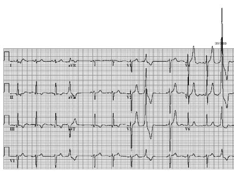
The extrasystoles have a right bundle branch block
morphology indicating an origin from the left ventricle.
The exact site of origin should be derived from the
information that comes from the extremity leads. There
is one extrasystole in the extremity leads exactly at the
moment that the leads change. Hence it should first be
decided whether the extra is recorded in leads I, II and
III or in aVR, aVL and aVF.
The clue is in the morphology of the extra in these leads.
A ventricular extrasystole can not be completely positive
in lead II and completely negative in lead III. The two
leads are 60° apart and concordantly the morphology
of the complexes in these leads is usually somewhat
similar. An opposite complex in aVL and aVF is well
possible and indicates an origin in the inferior wall. In
that case, the morphology of the ectopy in II and III
would also be expected to be negative. Indeed, this was
the case as shown in figure 2, which is the same ECG
printed in a different format.
Hence, the origin of the extras is in the inferior wall
and thus in the area of the old myocardial infarction. The
patient needs a serious work-up and an ICD should at
least be considered (MADIT II, SCD-Heft). In the
presence of nonsustained VTs on Holter monitoring
(and inducibility during EPS) a class I indication for
ICD implantation (according to the NVVC guidelines)
is established.

Latest revision as of 16:53, 8 October 2007

Author(s) A.A.M. Wilde, R.B.A. van den Brink
NHJ edition: 2005:2,67
These Rhythm Puzzles have been published in the Netherlands Heart Journal and are reproduced here under the prevailing creative commons license with permission from the publisher, Bohn Stafleu Van Loghum.
The ECG can be enlarged twice by clicking on the image and it's first enlargement
Figure 1

A 64-year-old man had an inferior myocardial infarction ten years ago. Lately he has been having palpitations with occasional dizziness. He sought the attention of his cardiologist. Physical examination revealed no particular abnormalities with the exception of a laterally displaced ictus cordis. His 12-lead ECG, shown in figure 1, was in sinus rhythm with some extrasystoles. The electrical axis is vertical and the Q waves and abnormal ST-T segments in the inferior leads are compatible with an old inferior myocardial infarction. An echocardiogram revealed reduced left ventricular function with extended inferior wall akinesia. The left ventricular ejection fraction was estimated to be 30%.

The questions to address are: what is the meaning of the extrasystoles, where do they come from and should further investigations be performed?

Answer