McGill Case 5: Difference between revisions
Jump to navigation
Jump to search
Created page with "{{McGillcase| |previouspage= McGill Case 1 |previousname= McGill Case 1 |nextpage= McGill Case 3 |nextname= McGill Case 3 }} [[File:E000583.jpg|thumb|600px|left|A patient wit..." |
No edit summary |
||
| (7 intermediate revisions by the same user not shown) | |||
| Line 1: | Line 1: | ||
{{McGillcase| | {{McGillcase| | ||
|previouspage= McGill Case | |previouspage= McGill Case 4 | ||
|previousname= McGill Case | |previousname= McGill Case 4 | ||
|nextpage= McGill Case | |nextpage= McGill Case 6 | ||
|nextname= McGill Case | |nextname= McGill Case 6 | ||
}} | }} | ||
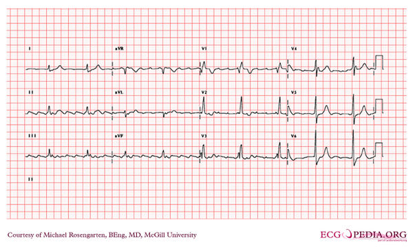
[[File: | [[File:E000705.jpg|thumb|600px|left|The rhythm is atrial flutter with flutter waves seen best in the inferior leads and in leads V1 to V3. The atrial rate is about 250/min. The QRS is wide (>120ms) an there is a tall R' wave in V1 and a shallow S in V6. The axis of the QRS seems normal. The EKG shows a right bundle branch block.]] | ||
Latest revision as of 05:08, 10 February 2012

|
