McGill Case 3: Difference between revisions
Jump to navigation
Jump to search
No edit summary |
No edit summary |
||
| (17 intermediate revisions by 2 users not shown) | |||
| Line 1: | Line 1: | ||
{{McGillcase| | {{McGillcase| | ||
|previouspage= | |previouspage= McGill_Case_2 | ||
|previousname= McGill Case | |previousname= McGill Case 2 | ||
|nextpage= | |nextpage= McGill_Case_4 | ||
|nextname= McGill Case | |nextname= McGill Case 4 | ||
}} | }} | ||
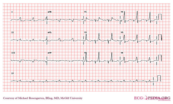
[[File: | [[File:E000703.jpg|thumb|600px|left|Intraventicular Conduction Defect | ||
comment: | |||
The ECG is in sinus rhythm and the QRS is markedly widened with a QRS duration of 260ms. The QRS seems split and gives the impression of ventricular bigemini but note that the second QRS deflection that looks like a PVC is in fact 200 ms after the onset of the first part of the QRS and hence too early for a PVC. Of interest this patient has recurrent ventricular tachycardia which may relate to his grossly widened QRS. | |||
The progression of the block can be seen over a three year period. | |||
comment from the web: | |||
When presented as a puzzler the correct interpretation of this ECG was not received, only suggestions of ventricular bigemini where given. | |||
]] | |||
Latest revision as of 05:06, 10 February 2012

|
