Answers: Difference between revisions
Jump to navigation
Jump to search
m Created page with '{{DRJcase| |previouspage= Case_reports_from_the_AMC |previousname= Return to list of cases |nextpage= DRJ2 |nextname= AMC case 2 }} Case 1|600px ==Questions=...' |
m →Answers |
||
| (3 intermediate revisions by 2 users not shown) | |||
| Line 11: | Line 11: | ||
==Answers== | ==Answers== | ||
# This ECG shows a severely prolonged QTc interval, which makes the patient prone to [[Torsade de Pointes]] and potential [[VF|ventricular fibrillation]] | # This ECG shows a severely prolonged QTc interval, which makes the patient prone to [[Torsade de Pointes]] and potential [[VF|ventricular fibrillation]] | ||
# Lead I has a negative P wave and Negative QRS complex. The arm leads were interchanged while recording this ECG. | # Lead I has a negative P wave and Negative QRS complex. The arm leads were interchanged while recording this ECG. | ||
# The S in v1 + the R in v5 (equaling 35mm or greater) are indicative of left ventricular hypertrophy | |||
Latest revision as of 05:41, 20 May 2010
This case report is kindly provided by Jonas de Jong and is part of the AMC case reports
|
Questions
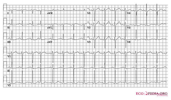
- This ECG was made shortly after this patient had been resuscitated. The patient was normothermic. What arrhythmia likely initiated the syncope?
- What technical abnormality is seen?
Answers
- This ECG shows a severely prolonged QTc interval, which makes the patient prone to Torsade de Pointes and potential ventricular fibrillation
- Lead I has a negative P wave and Negative QRS complex. The arm leads were interchanged while recording this ECG.
- The S in v1 + the R in v5 (equaling 35mm or greater) are indicative of left ventricular hypertrophy
