Puzzle 2009 08 Answer: Difference between revisions
Created page with 'ventricular fibrilation' |
m →Answer |
||
| (3 intermediate revisions by 2 users not shown) | |||
| Line 1: | Line 1: | ||
ventricular | {{NHJ| | ||
|mainauthor= '''A.A.M. Wilde''' | |||
|edition= 2009:08,307 | |||
}} | |||
[[File:Puzzle2009_08.jpg|thumb]] | |||
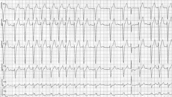
A 95-year-old woman presents with palpitations. She has no relevant medical history and the present | |||
complaint is several days old. She is not using any medication. Physical examination reveals an irregular | |||
pulse and heart beat (pulse deficit 20 beats/min) and no further abnormalities. Her ECG is shown in figure 1 | |||
(only the precordial leads are shown). | |||
'''How would you judge this ECG?''' | |||
==Answer== | |||
[[File:RhyrhmPuzzle2009_08_figure2.jpg|thumb|Figure 2]] | |||
The ECG (precordial leads) shows an irregular rhythm without clearly discernable P waves. Atrial fibrillation is the most likely supraventricular rhythm. QRS complexes show a left bundle branch block pattern (LBBB) with the exception of the last but 5th QRS complex. This complex is narrow and has a normal morphology. | |||
There are two potential explanations for this QRS complex. The first and probably most likely is that aberrant conduction occurs in the right bundle (concomitant conduction slowing in the contralateral bundle, the right bundle is contralateral to the left | |||
bundle that shows conduction delay in the other com- plexes) (figure 2). Indeed, aberrant conduction is most prevalent after the longest RR intervals during atrial fibrillation, as in this case. This relates to the longer refractory period of the bundles after a long preceding RR interval. The second possibility is ventricular ectopy in the ipsilateral bundle at the time that conduction passes through the right bundle. This possibility cannot be excluded. Ectopy should occur at the time that the supraventricular input reaches the site of the block. In both cases the QRS complex that ensues will mimic a normally conducted QRS complex. | |||
Latest revision as of 19:01, 23 October 2011
| Author(s) | A.A.M. Wilde | |
| NHJ edition: | 2009:08,307 | |
| These Rhythm Puzzles have been published in the Netherlands Heart Journal and are reproduced here under the prevailing creative commons license with permission from the publisher, Bohn Stafleu Van Loghum. | ||
| The ECG can be enlarged twice by clicking on the image and it's first enlargement | ||

A 95-year-old woman presents with palpitations. She has no relevant medical history and the present complaint is several days old. She is not using any medication. Physical examination reveals an irregular pulse and heart beat (pulse deficit 20 beats/min) and no further abnormalities. Her ECG is shown in figure 1 (only the precordial leads are shown).
How would you judge this ECG?
Answer

The ECG (precordial leads) shows an irregular rhythm without clearly discernable P waves. Atrial fibrillation is the most likely supraventricular rhythm. QRS complexes show a left bundle branch block pattern (LBBB) with the exception of the last but 5th QRS complex. This complex is narrow and has a normal morphology. There are two potential explanations for this QRS complex. The first and probably most likely is that aberrant conduction occurs in the right bundle (concomitant conduction slowing in the contralateral bundle, the right bundle is contralateral to the left bundle that shows conduction delay in the other com- plexes) (figure 2). Indeed, aberrant conduction is most prevalent after the longest RR intervals during atrial fibrillation, as in this case. This relates to the longer refractory period of the bundles after a long preceding RR interval. The second possibility is ventricular ectopy in the ipsilateral bundle at the time that conduction passes through the right bundle. This possibility cannot be excluded. Ectopy should occur at the time that the supraventricular input reaches the site of the block. In both cases the QRS complex that ensues will mimic a normally conducted QRS complex.