MI Diagnosis in RBBB: Difference between revisions
Jump to navigation
Jump to search
m MI diagnosis in RBBB moved to MI Diagnosis in RBBB |
mNo edit summary |
||
| Line 1: | Line 1: | ||
As shown in the examples below, [[Myocardial Infarction|myocardial infarction]] diagnosis in [[RBBB|right bundle branch block]] is not very different from normal MI diagnosis. | As shown in the examples below, [[Myocardial Infarction|myocardial infarction]] diagnosis in [[RBBB|right bundle branch block]] is not very different from normal MI diagnosis. As repolarisation in leads V1-V3 is often abnormal in RBBB, these leads cannot always be used for the diagnosis of ischemia. | ||
==Examples== | ==Examples== | ||
<gallery> | <gallery> | ||
Revision as of 08:51, 8 October 2009
As shown in the examples below, myocardial infarction diagnosis in right bundle branch block is not very different from normal MI diagnosis. As repolarisation in leads V1-V3 is often abnormal in RBBB, these leads cannot always be used for the diagnosis of ischemia.
Examples
-
 Patient with RBBB and inferior MI. Notice left axis deviation.
 -
 Lead V4R in the same patient with RBBB and inferior MI clearly shows ST elevation.
 -
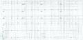 The same patient before acut MI developed. Horizontal axis.


