Clockwise and Counterclockwise rotation: Difference between revisions
No edit summary |
No edit summary |
||
| (One intermediate revision by the same user not shown) | |||
| Line 1: | Line 1: | ||
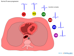
[[File:rwaveprogression.svg|thumb|right|300px|Normal R wave progression]] | |||
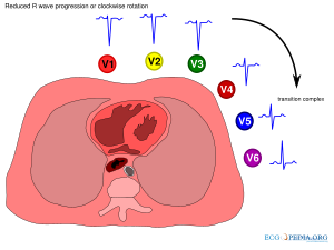
[[File:reduced_rwaveprogression.svg|thumb|right|300px|Reduced R wave progression or clockwise rotation]] | |||
Clockwise and counterclockwise rotation refer to a change in the electrical activity in a horizontal plane through the heart. Imaginge the observer standing at the feet of the patient who is in bed. If the electrical activity of the heart has turned more to the right side of the patient this is called ''counterclockwise rotation''. If the electrical activity of the heart has turned more to the left side of the patient this is called ''clockwise rotation''. | Clockwise and counterclockwise rotation refer to a change in the electrical activity in a horizontal plane through the heart. Imaginge the observer standing at the feet of the patient who is in bed. If the electrical activity of the heart has turned more to the right side of the patient this is called ''counterclockwise rotation''. If the electrical activity of the heart has turned more to the left side of the patient this is called ''clockwise rotation''. | ||
Latest revision as of 21:32, 14 July 2009


Clockwise and counterclockwise rotation refer to a change in the electrical activity in a horizontal plane through the heart. Imaginge the observer standing at the feet of the patient who is in bed. If the electrical activity of the heart has turned more to the right side of the patient this is called counterclockwise rotation. If the electrical activity of the heart has turned more to the left side of the patient this is called clockwise rotation.
Clockwise and counterclockwise rotation can be assessed only in the chest-leads (V1 - V6). Normally the R wave amplitude increases from V1 to V5. Around V3 or V4 the R waves become larger than the S waves and this is called the 'transitional zone'. If the transition occurs at or before V2, this is called counterclockwise rotation. If the transition occurs after V4, this is called clockwise rotation.
Several studies have been done to understand the anatomical substrate for these rotations. Tahara et al. have made CT-scans of 102 subjects to compare anatomic position of the cardiac septum and the electrocardiographic position of the transitional zone.Tahara Interestingly only 9 of 33 normal subjects had a normal transitional zone, which indicates that clockwise and counterclockwise rotation are not necessarily a sign of disease.
Causes of clockwise rotation were:
- intraventricular conduction abnormalities secondary to myocardial degeneration
- right ventricular heart disease
- shift of the septum to the left
- dilated cardiomyopathy
- shift of the whole heart
- pulmonary emphysema
- vertical heart (usually thin and tall persons)
Causes of counterclockwise rotation were:
- electrical shift to the right
- right ventricular hypertrophy
- WPW Syndrome
- Posterior myocardial infarction
- Left septal fascicular block
- shift of the septum to the right
- hypertrophic cardiomyopathy
References
<biblio>
- Tahara pmid=1919383
</biblio>