Answer MI 13: Difference between revisions
Jump to navigation
Jump to search
m New page: {{Case| |previouspage= MI 12 |previousname= MI 12 |nextpage=MI 14 |nextname=MI 14 }} '''Where is this myocardial infarction located?''' [[Image:ami0013.pg|700px|thumb|left|ECG MI 13. Clic... |
mNo edit summary |
||
| Line 7: | Line 7: | ||
'''Where is this myocardial infarction located?''' | '''Where is this myocardial infarction located?''' | ||
[[Image:ami0013. | [[Image:ami0013.jpg|700px|thumb|left|ECG MI 13. Click on image for enlargement.]] | ||
{{clr}} | {{clr}} | ||
==Answer== | ==Answer== | ||
Latest revision as of 10:47, 11 November 2008
| This page is part of Cases and Examples |
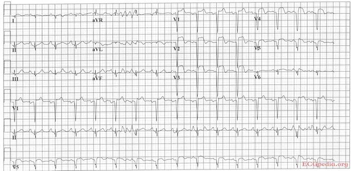
Where is this myocardial infarction located?

Answer
Culprit lesion: LAD
- sinustachycardia
- about 100/min
- normal conduction
- intermediate heart axis
- tall p wave in II consistent with right atrial dilatation. (the sawtooth-basline between the 2nd and 3rd complex in AVR is probably a motion artefact). PTA depression in II
- Loss of R waves throughout the anterior wall (V1-V6). QS complexes in V3-V5.
- ST elevation in V1-V5 with terminal negative T waves
- Conclusion: Large anterior MI due to LAD occlusion.
Characteristics that suggest a large infarct in this case are:
- Loss of R waves throughout the anterior wall (V1-V6). QS complexes in V3-V5.
- Left and right sided decompensation, resulting in right atrial dilatation and ischemia
- Tachycardia