Conduction: Difference between revisions
No edit summary |
No edit summary |
||
| Line 5: | Line 5: | ||
[[Image:QRSwaves.jpg|thumb]] | [[Image:QRSwaves.jpg|thumb]] | ||
The PQ time indicates how fast the action potential is transmitted through the AV node(atrioventricular) from the atria to the ventricles. Measurement should start at the beginning of the P wave to the beginning of the QRS segment. | The PQ time indicates how fast the action potential is transmitted through the AV node (atrioventricular) from the atria to the ventricles. Measurement should start at the beginning of the P wave to the beginning of the QRS segment. | ||
'''The normal PQ is between 0.12 and 0.20 seconds'''. | '''The normal PQ is between 0.12 and 0.20 seconds'''. | ||
A prolonged PQ time is a sign of a degradation of the | A prolonged PQ time is a sign of a degradation of the conduction system. This is called [[Arrhythmias #AV-block|1st, 2nd or 3rd degree AV block]]. | ||
A short PQ time can | A short PQ time can be seen in the [[Arrhythmias#WPW|WPW syndrome]] in which a faster connection exists between the atria and the ventricles. | ||
{{clr}} | {{clr}} | ||
==The QRS duration== | ==The QRS duration== | ||
The QRS duration indicates how | The QRS duration indicates how fast the ventricles depolarize. | ||
The ventricles depolarize normally within 0.10 seconds. When this is longer than 0.12 seconds, this is a '''[[ | The ventricles depolarize normally within 0.10 seconds. When this is longer than 0.12 seconds, this is a '''[[conduction delay| conduction delay (Left bundlebranchblock or Right bundlebranchblock]]'''. | ||
==The QT time== | ==The QT time== | ||
The QT time indicates how fast the ventricles are repolarized and how fast they are ready for a new | The QT time indicates how fast the ventricles are repolarized and how fast they are ready for a new heart cycle | ||
The | The normal value for QTc(orrected) is: 440ms for men and 450 ms for women. | ||
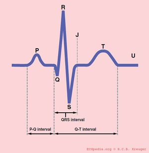
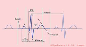
[[Image:QRSinterval.jpg|thumb| The QT interval start at the onset of the Q wave and ends where a line | [[Image:QRSinterval.jpg|thumb| The QT interval start at the onset of the Q wave and ends where a line for the steepest part of the T wave crosses the baseline of the ECG. Click on the image for a bigger image]] | ||
<flash>file=QTc.swf|width=200|height=135|quality=best|align=right|salign=R||bgcolor=#FFF5F5</flash> | <flash>file=QTc.swf|width=200|height=135|quality=best|align=right|salign=R||bgcolor=#FFF5F5</flash> | ||
The QT-interval comprises the QRS-complex, the ST-segment, and the T-wave. | |||
In a (serious) prolonged QT time, is takes longer for the myocardial cells to be ready for a new cardiac cycle. There is a possibility that some cells are not yet repolarized, but that a new cardiac cycle is already initiated. These cells are at risk for uncontrolled depolarization and induce a [[Arrhythmmias#Torsades_de_pointes|torsades de pointes]], a ventricular tachycardia. | |||
The QT interval is defined as follows: <cite>Lepeschkin</cite> The time between the beginning of the Q until the point where the steepest tangent line from the end of the T-wave intersects with the base line of the ECG. | |||
The difficult part is that the QT interval gets shorter if the heart rate increases. This cab be solved by correcting the QT time for heart rate using the Bazett formula:: | |||
[[Image:Formule_QTc.png]] | [[Image:Formule_QTc.png]] | ||
'' | ''at an RR interval 1 second, the (heart frequency 60/min) QTc=QT'' | ||
Using the QTc calculator on the right, the QTc is easy extractable. | |||
On the modern ECG machines, the QTc is given. However, the machines are not always capable of recognizing the correct QT time. Therefore, it is important to check this manually.. | |||
The following formula is indicative for normal values of QT time (uncorrected): | |||
[[Image:Formule_QTn_nl.png]] | [[Image:Formule_QTn_nl.png]] | ||
| Line 51: | Line 50: | ||
===Difficult QT times=== | ===Difficult QT times=== | ||
In some examples of the QT interval it can be difficult to measure a correct QT time. We have made a separate chapter: [[Difficult_QT| Measurement of difficult QT times]]. | |||
===Causes of | ===Causes of prolonged QT time=== | ||
* | *Medication (i.e. anti-arrhythmics, tricyclic antidepressants, phenothiazedes, for a complete list look on [http://www.torsades.org Torsades.org] | ||
* | *Inherited [[Long QT syndrome|long QT syndrome]] (LQTS) | ||
* | *Cerebral (subarachnoid haemorrhage, stroke, trauma) | ||
*Post infarct | *Post infarct | ||
===Short QT syndrome=== | ===Short QT syndrome=== | ||
There is also a rare form of the '''short QT syndrome''', in which the QTc < 300ms. This has been associated with sudden cardiac death.<cite>Gaita</cite> | |||
== References == | == References == | ||
Revision as of 13:56, 6 May 2007
Some statements may be disputed, incorrect or biased. |
The PQ time
The PQ time starts at the beginning of the atrial contraction and ends at the beginning of the ventricular contraction.
The PQ time indicates how fast the action potential is transmitted through the AV node (atrioventricular) from the atria to the ventricles. Measurement should start at the beginning of the P wave to the beginning of the QRS segment.
The normal PQ is between 0.12 and 0.20 seconds.
A prolonged PQ time is a sign of a degradation of the conduction system. This is called 1st, 2nd or 3rd degree AV block.
A short PQ time can be seen in the WPW syndrome in which a faster connection exists between the atria and the ventricles.
The QRS duration
The QRS duration indicates how fast the ventricles depolarize.
The ventricles depolarize normally within 0.10 seconds. When this is longer than 0.12 seconds, this is a conduction delay (Left bundlebranchblock or Right bundlebranchblock.
The QT time
The QT time indicates how fast the ventricles are repolarized and how fast they are ready for a new heart cycle
The normal value for QTc(orrected) is: 440ms for men and 450 ms for women.
<flash>file=QTc.swf|width=200|height=135|quality=best|align=right|salign=R||bgcolor=#FFF5F5</flash>
The QT-interval comprises the QRS-complex, the ST-segment, and the T-wave.
In a (serious) prolonged QT time, is takes longer for the myocardial cells to be ready for a new cardiac cycle. There is a possibility that some cells are not yet repolarized, but that a new cardiac cycle is already initiated. These cells are at risk for uncontrolled depolarization and induce a torsades de pointes, a ventricular tachycardia.
The QT interval is defined as follows: [1] The time between the beginning of the Q until the point where the steepest tangent line from the end of the T-wave intersects with the base line of the ECG.
The difficult part is that the QT interval gets shorter if the heart rate increases. This cab be solved by correcting the QT time for heart rate using the Bazett formula::
at an RR interval 1 second, the (heart frequency 60/min) QTc=QT
Using the QTc calculator on the right, the QTc is easy extractable.
On the modern ECG machines, the QTc is given. However, the machines are not always capable of recognizing the correct QT time. Therefore, it is important to check this manually..
The following formula is indicative for normal values of QT time (uncorrected):
Difficult QT times
In some examples of the QT interval it can be difficult to measure a correct QT time. We have made a separate chapter: Measurement of difficult QT times.
Causes of prolonged QT time
- Medication (i.e. anti-arrhythmics, tricyclic antidepressants, phenothiazedes, for a complete list look on Torsades.org
- Inherited long QT syndrome (LQTS)
- Cerebral (subarachnoid haemorrhage, stroke, trauma)
- Post infarct
Short QT syndrome
There is also a rare form of the short QT syndrome, in which the QTc < 300ms. This has been associated with sudden cardiac death.[2]
References
- LEPESCHKIN E and SURAWICZ B. The measurement of the Q-T interval of the electrocardiogram. Circulation. 1952 Sep;6(3):378-88. DOI:10.1161/01.cir.6.3.378 |
- Gaita F, Giustetto C, Bianchi F, Wolpert C, Schimpf R, Riccardi R, Grossi S, Richiardi E, and Borggrefe M. Short QT Syndrome: a familial cause of sudden death. Circulation. 2003 Aug 26;108(8):965-70. DOI:10.1161/01.CIR.0000085071.28695.C4 |
-
Bazett HC. An analysis of the time-relations of electrocardiograms. Heart 1920;7:353-370.

