McGill Case 356: Difference between revisions

From ECGpedia
Jump to navigation Jump to search
(Created page with " {{McGillcase| |previouspage= McGill Case 355 |previousname= McGill Case 355 |nextpage= McGill Case 357 |nextname= McGill Case 357 }} [[File:E356.jpg|thumb|600px|left|The rec...")
 
No edit summary
 
Line 7: Line 7:
}}
}}


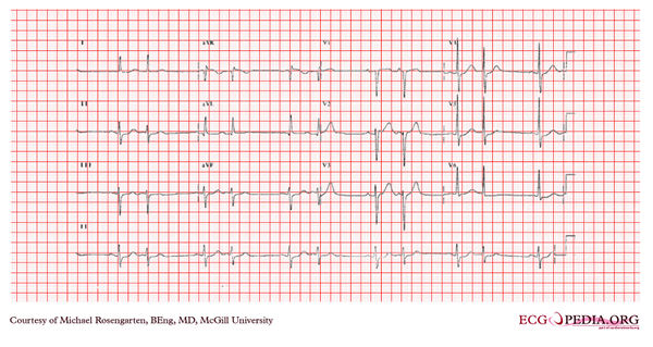
[[File:E356.jpg|thumb|600px|left|The recording shows sinus rhythm and a broad P wave in the inferior leads. The P wave is notched and the separation between the peaks is more than 40 ms. and the total p duration is more than 120 ms. This is suggestive of left atrial abnormality.
[[File:E356.jpg|thumb|600px|left|This is a regularly irregular rhythm at a rate of about 75/minute. There are two P wave morphologies best seen in lead V1. This is atrial bigemini.]]
The poor r wave progression in the precordial leads suggests a previous anterior wall myocardial infarction.]]

Latest revision as of 23:54, 19 February 2012

McGill logo.png
This case report is kindly provided by Michael Rosengarten from McGill and is part of the McGill Cases. These cases come from the McGill EKG World Encyclopedia.


Previous Case: McGill Case 355 | Next Case: McGill Case 357

This is a regularly irregular rhythm at a rate of about 75/minute. There are two P wave morphologies best seen in lead V1. This is atrial bigemini.